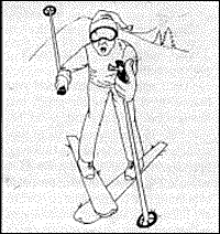
The typical history involves a patient who injures his thumb on the ski slopes; the patient may or may not remember a specific event. Physical examination will usually demonstrate instability in the first metacarpophalangeal joint, if the patient is examined before much swelling has occurred.
 Initially, radiographs may be negative, although small avulsed fragments from the base of the proximal phalanx can be delineated in some instances. These fragments may be displaced proximally and rotated from 45 to 90 degrees. Radiographs obtained with radial stress applied to the first metacarpophalangeal joint can reveal the luxation of the joint. It is important to stabilized this joint in order to prevent instability. This injury may take several months to heal and stress to the joint should be avoided.
Diagrams from Greenspan, A., Orthopedic Radiology, Gower Medical Publishing 1988, and Resnick and Naiwayama, Diagnosis of Bone and Joint Disorders, Sauders, 1981.
Click here for more information about Deborah Pate, DC, DACBR.


