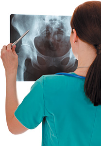
The 7th Interdisciplinary World Congress on Low Back and Pelvic Pain took place in Los Angeles, Nov. 9-12, 2010. This is a brief synopsis of some of the papers presented. There was a mix of scientific research and clinical research.
Standing Tolerance
Paul Marshall1 tested young people with no history of back pain and had them stand, unsupported, for two hours. (He stated that 30 minutes would have been enough for future trials.) The group divided into people with no pain on standing and people who developed pain on standing. The purpose; we suspect that people who develop pain when they stand are much more likely to end up with lower back pain. Pain developers were noted to have reduced side-bridge endurance. Men should be able to hold a side bridge for 83 seconds, women for 64 seconds. The other factor that correlated with pain development was gluteus medius co-activation, meaning that these patients co-contracted both gluteus medius when doing resisted hip abduction. Absolute strength and endurance of the gluteus medius did not correlate with pain developers.
Science is fascinating;, it does not always correlate with what we have been taught or with what we think is going to happen. This was one of the themes of the conference for me. You have to accept being confused by the science and remain open.
Treating the Ischial Spine for Pain in Pregnancy: More About Ligaments
 This was a fascinating study.2 The author took pregnant women with long-lasting sacral-area pain and did a relatively simple medical procedure. She injected steroids into the ischial spine, where the sacrospinous ligament inserts. The hypothesis: that problems with the pelvic ligaments contributed to pregnant women's pelvic pain. This injection gave long-lasting relief and changed not just local pain, but various referred pain patterns as well.
I have noticed that in many patients with sacral-area pain, some of the key spots are quite deep in the pelvis. I have spoken about friction to the ischial tuberosity, where the sacrotuberous ligament inserts.3 The ischial spine, where the sacrospinous ligament inserts, may be another important area to address with manual techniques.
Sacroiliac Diagnosis;4 Evidence-Based Physical Exam
Those of you who follow the research have probably noted that many of the usual tests chiropractors, osteopaths and manual therapists have used do not hold up; they are not consistent between examiners. There are a few tests that do seem to be reproducible and useful for physical exam of the sacroiliac. The study I am quoting compared patients with unilateral SI-area pain with normal, pain-free subjects. The tests with correlative positive findings included the following:
One: the active single-leg raise (ASLR). Interestingly, the result is based on the patient's subjective difficulty in lifting the straight leg. Two: the single-leg stance test. This was developed by Sahrmann and was further researched by Hungerford. Basically, you are testing the leg you stand on. As the patient lifts the opposite leg, you are visually observing the difference in motion between the PSIS and the sacrum. When the PSIS anteriorly rotates, it indicates an abnormal motion pattern, and does correlate with SI dysfunction and muscular dysfunction. (I described this test in detail in a previous article5.) Three: tenderness on palpation of the long dorsal SI ligaments. These ligaments can be easily palpated just below the PSIS, and extend medially down along the sacral border.
Are you using these three tests on patients you suspect of having SI pain? They are simple, they have literature support and they make sense. You can improve your evidence-based evaluation of the pelvis by including these tests in your evaluations.
Subgroups in Chronic Low Back Pain
There were several presentations on attempts to divide nonspecific chronic low back pain into subgroups. This is an ongoing topic in the research world, as clinical results for any therapy applied to the heterogeneous groups with nonspecific chronic lower back pain have not proven that effective. Can we find subgroups that enable us to target our patients more effectively? A broad-brush approach, attempting to define which group will respond to manipulation or a specific form of exercise, using clinical prediction rules, is one approach. Julie Fritz6 presented one of these approaches, while appreciating that it is not complete.
I was excited by a different approach to subgrouping. There were some fascinating presentations on chronic nonspecific lower back pain by a group of researchers who have worked in parallel over many years. These included Peter O'Sullivan,7 William Dankaerts8 and others. This group has looked at the biomechanics, trying to establish a pattern that underlies the pain. This is a broad approach, recognizing that all chronic lower back pain affects the brain and affects patients' behaviors. Clinical care has to be individualized for the specific patient.
They did not have simple single conclusion. Recognizing this complexity, they divided low back patients into four basic groups. A series of studies has reinforced this model, confirming it through both clinical tests of reproducibility by different examiners and scientific evidence, including EMG studies, of the nature of these muscle imbalance patterns.
The first two groups involve a smaller percentage of patients. The first includes those who have a true anatomic pathology driving their pain. As much as imaging overclassifies patients into this category, there are some folks whose pathology is the main ongoing pain generator. The second group includes those who primarily have a biopsychosocial problem, whose problem is mostly about psychological behavior and maladaptation to their initial pain.
The third and fourth categories are larger. The third group was fascinating to me. The authors call it a movement impairment classification, which means these patients have stopped moving in a particular direction and compensate by overactivating the antagonists to this motion. These folks typically start with a severe lower back episode often involving flexion and were trained, either by their initial pain or by some well-meaning therapist, to absolutely avoid a particular motion; often to avoid flexion. They then compensate by overactivating the spinal extensor muscles to avoid that direction, and end up with a fear-based behavior driving their spinal function. They have excessive spinal stability and increased loading of the spine due to muscular co-contraction of the abs and the back muscles.
The guarding itself creates huge loads on the spine. (I suspect I have been guilty of creating a few of these types myself.) Therapy for these patients focuses to some degree on cognitive behavioral therapy, working through their fear, which is the first step in normalizing their movement pattern. They need to relax, they need to gradually learn how to bend forward freely, and they need to let go of their chronically tight muscles.
In my fascination with stability, I wonder how many of these patients I have either not noticed or have mistreated. I wonder how many of the patients who end up getting facet ablation from the injection docs are really in this category; and are just overloading their facets through holding themselves in extension and overfiring their lumbar extensors.
The fourth category is one of control impairment. These folks have minimal to no awareness of their spinal positioning. They typically fail to control a motion, often flexion, sometimes extension, sometimes multiple movements, which reproduces their symptoms. They cannot control the symptomatic area of the spine in the direction that recreates their symptoms. They may have tended to have a more gradual onset of pain; thus they can lack a withdrawal reflex from the pain, and this is reinforced by their lack of proprioceptive awareness. These are the patients who will respond to a stabilization strategy.
Visceral Manipulation: The Kidney
Another study that was fascinating to me involved the effect of visceral manipulation of the kidney on kidney motion and low back pain. Clinically, I have used and appreciated visceral manipulation for many years, but there has been minimal scientific evidence of its utility. Paolo Tozzi9 used ultrasound imaging to record the motion of the kidney during breathing and then see if it would change after manipulation. This was an elegant study that opens doors for further research.
The Hip Joint
There were several presentations about the hip joint. Patients with hip DJD and other hip problems are very likely to have concurrent low back and/or pelvic pain. Recent research has noted that lower back patients with a lack of motion of the hip, especially a lack of internal rotation, do not respond as well to low back manipulation. Hip labral tears were discussed.10 One of the physiatrists who presented had a novel diagnostic triage for this. First, she injected the hip joint itself with local anesthetic to see if doing so would change the pain pattern. This was done before any MRI imaging of the hip, as a screening. The question: Is the hip joint a major cause or contributor to the hip, lower back and/or pelvic pain? She noted that most patients with hip labral tears are not candidates for surgery, but rather need physical medicine and rehab first.
This was a great conference. My review just scratches the surface of the many presentations. It was wonderful to see and hear some of the top names in back research from multiple different professions. The ongoing tension and dialog between clinicians and researchers was apparent. We all can learn from each other. As a profession, we as chiropractors need to increase our appreciation and utilization of the most current evidence.
Author's note: Most of the references below and enumerated in the text refer to presentations given during the World Congress.
References
- Paul Marshall. "Hip Muscle Strength, Endurance and Co-Activation as Predictors of Low Back Pain During Prolonged Standing."
- Anne Lindgren. "Increased Physical Function After Locally Administered Corticosteroid to the Ischiadic Spine. A Randomized Double Blind Controlled Trial on Women With Persistent Pregnancy-Related Pelvic Girdle Pain."
- Heller M. "Sacroiliac Revisited: The Importance of Ligamentous Integrity." Dynamic Chiropractic, July 2, 2005.
- Tiina Lahtinen-Suopauki. "Association Between Rotational Movement Control Dysfunction of the Pelvis in One Leg Stance, Positive Scoring in Active Straight Leg Raise Test and Tenderness in the Dorsal Sacroiliac Ligament."
- Heller M. "Stabilizing the Pelvis With Exercise; (Relatively) Simple Rehab Strategies." Dynamic Chiropractic, March 26, 2009.
- Julie Fritz. "Identifying Subgroups of Patients Within Physiotherapy."
- Peter O'Sullivan. "Exercise and Back Pain - What Type, How and for Whom?"
- Wim Dankaerts. "Identifying Subgroups of Patients From a Biomechanical Perspective."
- Paolo Tozzi. "Evidence-Based Correlation Between Low Back Pain and Reduction of Renal Mobility, Assessed by Dynamic Ultrasound Topographic Anatomy Evaluation (D.U.S.T.A.-E.): Local Kidney Manipulation Improves Kidney Mobility and Decreases Pain Perception."
- Heidi Prather. "The Better You Are, the Greater the Risk: Hip Findings in Female Soccer Players Vary With Level of Experience."
Click here for more information about Marc Heller, DC.


