


In my previous two articles, I began discussing a protocol for adjustment of the pronated foot. I will conclude with:
Associated Pronation Adjustments
#1. Hallux Valgus (hallux = toe. valgus = deviation away from mid-line). This condition eventually leads to a bunion, which is a callus over an inflamed bursa.
Adjustment: The doctor stabilizes the lateral border of the foot with the "outside" hand while the "inside" hand index finger contacts the medial border of the foot with the thumb over the shaft of the "big" toe. The thrust is a pull toward the doctor. No varus/medial thrust is given - this may cause a spraining of the capsule. With a symptomatic patient, the doctor should lighten the "inside" hand contact and release contact at the end of the pull.
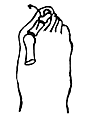
Figure 1: A hallux valgus condition.
#2. Heel Spur Adjustment. This is a very basic adjustment that attempts to approximate the heel to the toes.
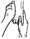
Figure 2: The hallux valgus adjustment.
Adjustment: The patient is prone with the knee bent at 90 degrees. The doctor stands at the outside of the involved foot. The headward hand "cups" the calcaneus with the thenar as the footward hand grasps the forefoot. The headward hand thrusts toward the footward hand, and vice versa.
#3. Toe Mobilization. This is an excellent mobilization procedure. The doctor's thumb pad contacts the shaft of the toe while the distal interphalangeal joint of the index finger contacts the plantar surface of the proximal or distal interphalangeal joint. The "outside" hand stabilizes lateral aspect of the foot. The doctor utilizes traction thrust preceding by one or two rotations.
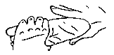
Figure 3: The heel spur adjustment.
#4. Metatarsal Mobilization. This procedure is for general mobilization of the midfoot and forefoot. The doctor grasps the dorsum of foot with the thenars (thumb pointed toward knee) and finger pads on plantar surface. The doctor performs opposing "figure-eight" motions to induce motion into the foot.
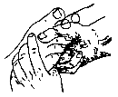
Figure 4: Toe mobilization.
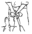
Figure 5: Metatarsal mobilization.
Conclusion
Excessive pronation is associated with many musculoskeletal complaints, from the foot itself, up the leg to the knee, hip, and even the pelvis and spine. The good news is that all of these conditions can be helped with custom-fitted orthotics. Investigation of foot biomechanics is a good idea in all patients, but especially for those who are recreationally active. Many times, correction of recurring subluxations can only be accomplished when an excessively pronating foot is provided with appropriate orthotic support.
Mark Charrette,DC
Las Vegas, Nevada
Click here for previous articles by Mark Charrette, DC.