


To evaluate for weak abdominals, have the patient lie supine with the legs extended and the arms behind the neck. Ask the patient to lift the legs upward. If the abdominal muscles are strong, the back can be held flat on the floor by the abdominals, holding the pelvis in posterior tilt position during the leg-raising movement. If the abdominal muscles are weak (Figure 1), the pelvis tilts anteriorly as the legs are lifted and the back will hyperextend into lordosis.2 Put your hand in the lumbar area before the test to feel for a possible abnormal lordotic change or normal spinal flattening against the table. Abdominals may also be considered normal (grade 5) when a supine patient (with hands clasped behind the head, with the chin tucked) can bring their head, shoulders and arms off the table as in a sit-up until the inferior angles of the scapulae are off the table.3
Figure 1: Abdominal muscle test. From Kendall FP, McCrearly EK, Provance PG. Muscles Testing & Function 3rd ed. Baltimore, MD. Williams & Wilkins:137,1993; with permission. |
A simple test for shortening of the lumbar erector spinae fascia is for the seated patient, knees over the end of the table, to first "fix" the pelvis by the patient placing his hands on the iliac crests and bending the neck and spine forward to "hump" the lumbar spine. If the lumbar fascial section of the spine is shortened no lumbar kyphosis will occur.4 Another method is to have the seated patient bend the neck forward followed by the rest of the spine, while the practitioner's hands are placed on the crest of the patient's ileum. The patient's head should reach within eight inches of the femur before the iliac crests move. Lewit states that if the patient has a short trunk and long thighs, the test may create a false negative; if the patient has a long trunk and short thighs, a false positive short erector spinae may occur.3
Another basic way of determining where to treat the restricted posterior thoracolumbar fascia, besides palpating for fascial barriers, is to have the patient flex forward in both the sitting and standing positions. It has been found that there is increased erector spinae pressure (decreased blood flow) in vivo in both normal and spinal patients in the flexed position while standing and with loading.5 The patient is also asked to flex forward in rotation right and left from the cervical spine to the lumbar spine, and in lateral bending. Ask patients where they feel strain, tension or pain on these movements, and palpate these areas for fascial barriers. After treatment, these same movements can be re-evaluated to determine areas that persist, or new areas of involvement.
Effective methods of treating restricted posterior spinal fascia can be accomplished by fascial release (Figure 2). In this method, after the restricted area is found by flexion testing and palpation, the doctor contacts the area in the direction of the barrier and waits for the fascia to release. If the release does not occur within 30 seconds, ask the patient to inhale for 10 or more seconds, which often enhances the release of the tissue. The doctor then follows the tissue to the end, or waits at a new barrier, if present, to continue the release. As the tissue releases, the doctor can move the patient in a direction that stretches the direction of the release of the barrier.6
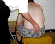
Figure 2: The right hand is directed caudally into the barrier. From Kendall FP, McCrearly EK, Provance PG. Muscles Testing & Function 3rd ed. Baltimore, MD. Williams & Wilkins:137,1993; with permission. |
Another method of release involves the use of Graston instruments. Figure 3 depicts the use of the Graston handlebar instrument over the lateral lumbar fascia. Before the handlebar was used to release the fascial restriction, this instrument or other Graston instruments were used to first confirm the area of fascial restriction and the direction of the fascial barrier.7
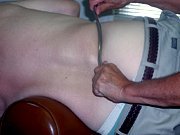
Figure 3: The Graston method for lateral lumbar fascia in the direction of the barrier. From Kendall FP, McCrearly EK, Provance PG. Muscles Testing & Function 3rd ed. Baltimore, MD. Williams & Wilkins:137,1993; with permission. |
References
- Solomon R, Brown T, Gerbino PG, Micheli LJ. The young dancer. Clinics in Sports Med 2000;19(4):717-739.
- Kendall FP, McCreary EK, Provance PG. Muscles: Testing & Function, 4th ed., Baltimore, Williams & Wilkins,1993:137.
- Hislop HJ, Montgomery J. Muscle Testing. Philadelphia, WB Saunders Co., 6th ed., 1984.
- Lewit K. Manipulative Therapy in Rehabilitation of the Locomotor System. Boston, Butterworth Heinemann. 3rd ed., 1999.
- Konno S, Kikuchi S, Yoshihiro N. The relationship between intramuscular pressure of the paraspinal muscles and low back pain. Spine 1994;19(19):2186-9.
- Hammer WI. Genitofemoral entrapment using integrative fascial release (IFR) Chiropractic Technique 1998;10(4):169-176.
- Hammer WI. Graston - A necessary part of the puzzle. Dynamic Chiropractic 2001;19(20).
Warren Hammer,MS,DC,DABCO
Norwalk, Connecticut
Click here for previous articles by Warren Hammer, MS, DC, DABCO.