


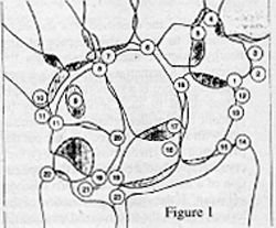
Key to Figure 1:
- epitrapezium
- calcification (bursa or flexor carpi radialis)
- paratrapezium
- secondary trapezium
- secondary trapezoid
- os styloideum
- Gruber ossicle
- secondary capitate
- os hamuli proprium
- os versalianum
- os ulare externum (bursal or tendinous calcifications)
- os radiale esternum
- traumatic avulsions
- unfused ossification center of the radial styloid process
- intercalary ossicle between scaphoid and radius (os paranaviculare)
- os centrale carpi
- hypolunate
- epilunate
- accessory ossicle between lunate and triquetrum
- epipyramis
- triangulare
- unfused ossification center of the ulnar styloid process
- small ossicle at the radioulnar articulation
Figure 1 demonstrates the potentially visible accessory bones that can be present on a PA view of the wrist. Many ossicles probably correspond to a capsular ossification as a manifestation of degenerative changes rather than a true anomaly. The epitrapezium may represent bursal calcification or tendon calcification. The hook of the hamate ossifies independently and if it does not fuse, it remains isolated and is seen as an accessory bone, the os hamuli proprium. When seen in the lateral view it can be mistaken for a fracture. The os ulnare externum can be an acquired ossification or calcification and should be considered in the differential diagnosis. A separate ossification center can occur at the radial styloid process persisting to imitate a super numerary skeletal element. Along the ulnar aspect of the styloid process, a small ossicle is occasionally found. Between the scaphoid and the articulating surface of the radial styloid process. This may also be ossification in the tendon or tendon sheath.
The most important factor is to determine if the ossicle is due to recent trauma or if it represents an old degenerative change or an anomaly. If there is a question as to the most accurate diagnosis, take an x-ray of the opposite wrist. With an anomaly frequently they are bilateral. If there is still a question, repeat the study in 14 to 21 days. If it is a recent injury there should be evidence of healing present on the later study indicating the accessory element was a recent injury.
Reference
1. Kohler and Zimmer: Borderlands of Normal and Early Pathologic Findings in Skeletal Radiology. Thieme, 1993.
Deborah Pate, DC, DACBR
San Diego, California
Click here for more information about Deborah Pate, DC, DACBR.