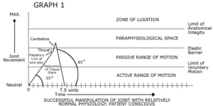
Graph #1: This illustration of a high-velocity chiropractic adjustment is an example of the Diversified/Palmer/Gonstead adjustments which are usually effective in approximately 95 percent of our patients where adjustment/manipulation is the treatment of choice. The graph consists of a vertical "ordinate" representing a single vector of joint movement/distraction ranging from the joint's neutral position through its active range of motion, continuing along the vector through passive range of motion, past the elastic barrier, and into the paraphysiological space.3 At the top of the chart, a line marked maximum illustrates the limit of anatomical integrity and any movement past that would cause a dislocation of the articulation. Horizontally along the bottom of the graph, the "abscissa" is a function of time with nondescriptive time units.
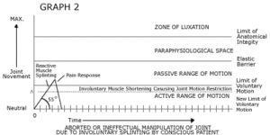
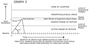
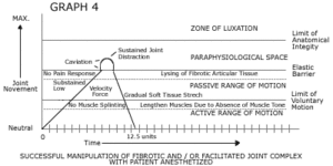
The graphs are designed to be conceptual and qualitative, not quantitative. There are no standard units used of joint distraction, slope of force and velocity, nor of units of time.
On graph number one, the slope of the force and velocity applied in the manipulation is represented by a diagonal line coming from the intersection of the graph illustrating neutral joint position and zero times rising at a 55 degree angle which is an arbitrary relative illustration. Follow the diagonal line representing joint movement passing through the active range of motion, into the passive range of motion, and reducing tissue slack to the point of the palpatory limit of joint "end-play." At that point, the skilled chiropractor will deliver a high-velocity thrust represented as an 85 degree slope of velocity toward and through the elastic barrier.
Once past the elastic barrier, an instantaneous negative barometric pressure is achieved within the closed joint structure causing a dissolution of gasses in the fluids to form bubbles causing the audible "click" or cavitation.4
With this instantaneous loss of a relative vacuum, the "potential space" within the joint is then converted to an "actual space" allow greater movement and stretching of the articular soft tissues. The momentum of the high-velocity thrust may continue the joint separation past the point of cavitation, at which time the resiliency of the capsule again approximates the apposing articular beds and the joint is then allowed to seek its own juxtaposition, initiating a change in physiology and function. The descending diagonal line would illustrate the time involved for the tissues to return to their resting length and re-establishment of resting tonus. The adjustment illustrated on Graph 1 would be effective in the normalization of joint movement in the majority of cases where chiropractic manipulation is indicated. This application could and would stretch shortened tissues and disrupt adhesions in the joint complex.
Graph #2: This illustrates the minority of situations where joint manipulation is indicated but where fibromuscular indurations have formed as a result of trauma, chronic spasm, or the effects of immobilization have caused a restriction in active range of motion. Many times when we attempt to adjust the joints in the presence of this aberrant muscle function, we encounter an inability to guide the joint through the active range of motion and into the passive, thus not achieving movement enough to detect joint end-play which would allow us to deliver an effective chiropractic adjustment. Many times this resistance is due to reactive muscle splinting and pain response by the patient resulting in an aborted or ineffectual manipulation of this joint due to the conscious patient's response to the manipulation.
Graph #3: This illustrates the minority of cases where rigid fibrosis of the normally elastic supportive tissues impedes our ability to pass through the passive range of motion resulting in reflex protective splinting or in a pain response. When a thrust is applied, there may even be some tearing of fibrotic tissue, but the force is unable to pass effectively through the fibrotic blockage and then through the elastic barrier enabling us to effectively distract the joint into the paraphysiological space.
Graph #4: This is an illustration of manipulation with the patient anesthetized. The slope of the diagonal line is demonstrated as being at a 45 degree angle, again an arbitrary and qualitative illustration indicating the low-velocity technique. Careful application of force into the articulation allows the moving articulation to pass through the aberrant muscle tone, stretching the shortened muscle past active range, through the passive range to where the manipulator would palpate the fibrotic scar tissue resistance, continuing through and lysing those fibrotic tissues to the elastic barrier and applying consistent low-velocity/high amplitude movement into the paraphysiological space. At this point, the decreased intra-articular barometric pressure will cause a cavitation of the joint. The joint literally "falls" open due to the change of the relative vacuum within the joint. A brief continuation of force is then applied stretching the capsule and lysing any intra-articular adhesions and fibrosis. The force is discontinued and the tissues return to their resting state. A successful manipulation of the motor unit has been accomplished which serves two functions: 1) stretching of supportive tissues, and (2) the lysing of fibrotic adhesions and joint blockages.
There is an additional minimum amount of distraction in the paraphysiological space that is achieved with the MUA patient as compared to the conscious patient but not to the point where it would breach the limit of anatomical integrity. If you will notice, the cavitation which is achieved in the conscious patient is a result of a high-velocity thrust applied towards the end of the passive range of motion through the elastic barrier, and is merely incidental to the high-velocity thrust that is applied. The cavitation in the anesthetized patient itself causes the rapid distraction of the joint due to the articular surfaces basically "falling open."
These graphs are effective in illustrating the concept of restoration of movement through the chiropractic adjustment, both in the conscious and anesthetized patient.
MUA as taught by the pioneer of the training programs, Stephen Capps, D.C., involves two aspects: 1) the passive stretching of large muscle groups affecting gross biomechanics, and 2) the specific attention given to dysfunctioning motor units. These graphs are directed toward the latter. Any practitioners familiar with proprioceptive neurofacilitation technique and comparable muscle stretching techniques such as strain/counterstrain and muscle energy would appreciate the importance of addressing the shortening of the larger muscle groups in the restoration of spinal function.
For further information regarding MUA, contact Tim Mills, D.C., c/o MUA Associates of Southern California, P.O, Box 16305, Beverly Hills, California 90209-2305, (310) 864-3627.
References:
- Greenman, PE: Manipulation with the patient under anesthesia. J. Amer. Osteopathic Assoc., 92(9):1159-1167, Sept. 1992.
- Faye, LJ and Schafer RC: Motion Palpation and Chiropractic Technic, ed 2. MPI 1991.
- Sandoz R: Some physical mechanisms and effects of spinal adjustments. Ann. Swiss Chiro. Assoc., 6:91, 1976.
- Cyriax JH: Orthopedic Medicine Volume Two -- Treatment By Manipulation, Massage, and Injection.
Timothy L. Mills, DC
Cypress, California


