


Radiation therapy (RT) is an important modality in the treatment of a wide variety of neoplasms. The complications associated with radiation therapy to the skeletal system include alterations in bone growth, radiation osteitis with secondary stress fractures, osteonecrosis, infection as well as radiation-induced sarcoma.
This is a very generalized overview of the radiographic features of radiation-induced changes in bone that one might encounter in our chiropractic patient population. The patient's history is extremely important to determine whether the radiographic findings are related to previous treatment. I am only reviewing the most common findings in order to allow you to distinguish these changes from other pathological conditions.
I also do not want the reader to assume all patients who are treated with RT ultimately will end up developing osteoradionecrosis and malignant neoplasms. This is simply a brief overview to remind the clinician that a patient with a history of neoplasm treated with RT may demonstrate osseous changes.
The main effects RT has upon bone are disruption of normal growth and maturation, scoliosis, osteonecrosis and neoplasm formation. Growth arrest is believed to be caused by a combination of cellular injury to chondrocytes and damage to the blood vessels. The epiphysis is the most sensitive area to radiation. Radiation therapy has also been shown to increase the risk for epiphyseal plate injury such as a slipped capital femoral epiphysis. RT delivered to the metaphysis can cause bowing, fraying and the appearance of sclerosis, similar to the changes found in rickets. The diaphysis is thought to be relatively radioresistant and periosteal bone formation may be less affected than enchondral bone formation. However, when an entire bone isexposed to radiation, it will demonstratenarrowing of the shaft and an overall reduction in size and diameter. A common complication with RT is scoliosis, which affects the immature spine more severely than a mature spine.
Complications in the mature skeleton include osteonecrosis, pathologic fracture and radiation-induced neoplasms. The following (Table 1) is a generalized chart of the bone changes and time period for bone that has been radiated. Please keep in mind that these changes do not occur in every case and they are dependent on dosage, associated treatments, age and general health of the patient and a multitude of other factors. This is only a very brief outline to give a simple guideline as to when changes might occur.
Table 1
|
Radiation necrosis is dose dependent. Radiation osteitis is a term used to describe potentially reversible changes such as temporary cessation of growth, periostitis, bone sclerosis and increased fragility, ischemic necrosis and infection. On radiographs, the bone will appear mottled, demonstrating both osteopenia and sclerosis and areas of coarse trabeculation. Changes are seen at different time intervals depending on which bone is irradiated. The following is a list of bones commonly affected, along with their radiographic characteristic findings.
- Mandible: Demonstrates the radiographic features of osteoradionecrosis (defined as irradiated bone that fails to heal over a period of three months), which in the mandible appears as ill-defined cortical destruction without sequestration. This complication, which is relatively rare, generally is seen within a year of treatment. Unfortunately, being a superficial bone, it inadvertently receives a larger dose of radiation during treatment of head and neck neoplasms. Differentiating osteoradionecrosis from tumor recurrence can be very difficult.
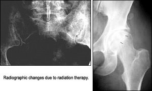
- Pelvis: Avascular necrosis of the femoral head can occur; in a very small percentage of patients, fractures of the femoral head can occur.The fractures generally heal with normal callous formation. The SI joints also can be affected, demonstrating bilateral or unilateral sclerosis depending on the field of radiation. The sacrum also can be affected similarly.
- Shoulder: The shoulder frequently receives radiation in the treatment of breast carcinoma. This can induce osteopenia and/or a coarse and disorganized pattern of trabeculation, mimicking Paget's disease. Rib, clavicular and scapular fractures also can occur and in some cases; avascular necrosis of the humeral head can develop.
- Sternum: Osteopenia, disorganized trabeculae and necrosis involving one or more sternal segments can occur, with associated pectus excavatum.
- Spine: Radiation-induced changes of the spine are best revealed with MR. The hematopoietic cellular elements of the marrow are replaced with fat, which is bright on T1-weighted sequences. Osteopenia and associated compression fractures can occur.
Radiation-Induced Neoplasms
Radiation-induced tumors in patients under the age of two years are most often benign, with osteochondromas being the most common. In the case of adults, however, this is not always true case. A late effect of ionizing radiation is the development of sarcoma within the field of irradiation, referred to as postradiation sarcoma (PRS). The overall incidence of PRS is less than 1 percent for patients with cancer who are treated with radiation and survive five years.1 Although the implication for individual patients is significant, there is little doubt that the benefits of ionizing radiation outweigh the potential risks for developing sarcomas.
References/Resources
- Inoue YZ, Frassica FJ, Sim FH, et al. Clinicopathologic features and treatment of postirradiation sarcoma of bone and soft tissue. J Surg Oncol 2000 Sep;75(1):42-50.
- Mitchell, MJ, Logan, PM. Radiation-induced changes inbone, Journal Radiographics 1998;18(5):1125-1136
Click here for more information about Deborah Pate, DC, DACBR.