


Patellofemoral pain syndrome (PFPS) is a common cause of knee pain.1 In fact, it is the most often diagnosed disease of the knee in orthopaedic and physiotherapy clinics.2 Once it starts, PFPS becomes chronic and seriously limits physical activities in adults.3 Surgery for PFPS is costly and has not been shown to be useful.4
Etiology and Diagnosis
Certain authors theorize that PFPS is caused by a degeneracy of the cartilage, but it is well-known that cartilage has no nerve cells, and that many people have no pain even if the cartilage is substantially altered, while others have a lot of pain with normal cartilage.
The patellar grinding test is considered to be the most specific for PFPS.8-9 It is done with the patient's leg in extension; the examiner grasps the patella with one hand on the other, and applies a compressive pressure on the patella vertically into the patellofemoral joint space.5, 9-10 The test is positive if it causes pain.
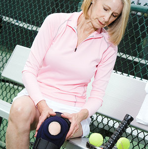 The grinding test has to be performed delicately at the beginning, gradually augmenting pressure to the patient's tolerance. The greater the pain, the more serious the condition. This test is frequently positive even in asymptomatic patients; it is only past a certain level of irritation that the symptoms appear.
The most common causes of PFPS are overwork, bad alignment of the patella and trauma.11-12 For example, an office worker decides to redo a wooden floor. For three days in a row, they kneel and/or squat, and a few days later the symptoms appear.
Treating PFPS With Ischemic Compression
The present author carried out a randomized trial on PFPS.13 The aim of this trial was to test the efficacy of 15 myofascial therapy treatments using ischemic compressions directly to the knee in 38 patients suffering from chronic PFPS. The experimental group (27 patients) received treatment directly to the knee, while the control group (11 patients), received ischemic compressions to the trigger points (TrPs) localized in the hip muscles.
The patellar grinding test was used with a scale from 0-5 to quantify the pain present; the test was performed by another chiropractor who did not participate in the treatments.
Intervention: When the TrPs were located under the patella, treatment was like the examination: the patella was crushed in the hyperirritable foci (1 or 2); the pressure was very light at the beginning and gradually increased to patient tolerance. Then the pressure was kept up for 8 seconds without moving.
When TrPs were found (one or two) at the peripatellar level, they were treated with thumbtip pressures in a 90 degree angle. The 8-second pressure was to patient tolerance and repeated at each visit until elimination, or in this trial, within 15 treatments. In all cases of PFPS, there are TrPs under the patella (retropatellar), but not necessarily at the plica level (peripatellar).
My hypothesis is that passive and oriented mobilization of the patella was an ischemic compression on the TrPs located under the patella. The ones at the peripatellar level were treated directly with thumbtip pressure. TrPs may be located in muscles, ligaments, tendons, fasci and articular capsules.14
Results of this trial demonstrated a decrease in the irritation with the patellar grinding test. After 15 treatments, the treatment success rate was 60 percent in the experimental group versus 28 percent in the control group. In the experimental group, there was still a 43 percent amelioration six months later.
The aim of these treatments should be the complete elimination of all the TrPs. Often, right after the first few treatments, there is great improvement. As well as patellofemoral pain syndrome, many patients have TrPs somewhere around the knee, so it is best to treat all the problematic areas, especially the ones pinpointed by the patient. At the site indicated, every square centimeter should be examined attentively with thumbtip pressure.
Some patients have edema at the anterior part of the knee; normally, this will vanish while taking care of the PFPS. In obese patients, the knee may be deformed with calcium deposits and hyperirritation, mostly at the interior part of the knee (osteoarthritis), at the level of the meniscus and at the lower insertion of the vastus lateralis and medialis muscles. All these irritations can be treated with ischemic compressions and these patients, even those with major problems, can be significantly helped. A good way to treat the inside of the knee is to have the patient lie on the symptomatic side while you apply an 8-second thumbtip pressure vertically on each TrP.
Pain at the interior or exterior meniscus, with or without tears, can be treated with very good results using ischemic compressions. Examination with magnetic resonance has shown that 67 percent of people over age 65 have tears of the meniscus without any symptoms.15 Surgery may be warranted when there is a tear plus misplacement of the meniscus and the knee cannot be extended.
Epicondylitis: Overview and Treatment
Lateral epicondylitis, also known as tennis elbow, is the most common elbow problem in adults. Normally it is produced by an accumulation of microtraumas causing inflammation and degeneracy of the musculotendinous tissue attached to the epicondyle. This problem may be extremely debilitating, even with regard to the performance of normal daily activities.16-17
Occupational activities that necessitate repeated forced dorsiflexion of the forearm, and jobs involving radial deviation with the arm in supination, such as plumbing and computer work, may cause stress at the extensor muscles of the wrist. This can result in degenerative changes to the common tendon of these extensors at their point of attachment on the lateral epicondyle of the humerus. As the name suggests, is often seen in tennis players.18-19
In a trial by Hay20 on epicondylitis, four weeks of anti-inflammatory medication proved no more effective than placebo. For Ollivierre,16 cortisone injections may attenuate the symptoms, but they cause degeneracy, weakness and sometime a rupture of the musculotendinous unit. Surgery may be useful in less than 5 percent of cases of lateral epicondylitis.21
The treatment I use with this condition is as follows: With the patient supine, stretch their arm along their body, applying an 8 second thumbtip pressure on the trigger points (TrPs) localized along the extensors muscles and at the epicondylitis. These TrPs may be very sensitive, so you should proceed delicately at the beginning, gradually augmenting the pressure to the patient's tolerance. The extensor muscles are attached to the epicondyle and descend for 8 centimeters along the radius.
Another approach may be used concurrent to the aforementioned treatment. The TrPs may be treated with the patient kneeling on the floor, perpendicular to the table, their forehead resting on the asymptomatic arm. Stretch the arm along the table with the hand in pronation. The TrPs may then be reached easily; an 8-second pressure on each will normally produce evident amelioration within six treatments, in my experience.
In cases of medial epicondylitis, the irritation is on the inside of the humerus. The TrP may be reached with the patient's hand supine.
Every patient has some irritation at these sites, but the symptoms will appear only above a certain level. Sometimes it is useful to check for pain when you press the elbow directly from posterior to anterior. If this mobilization produces pain, an 8-second pressure, repeated until the hyperirritability is gone, may affect the overall elbow function. Chronic medial or lateral epicondylitis may need 10-20 treatments before resolving.
References
- Crossley K, Bennell K, Green S, McConnell J. A systematic review of physical interventions for patellofemoral pain syndrome. Clin J Sport Med, 2001;103-10.
- McMullun W, Roncarati A, Koval P. Static and isokinetic treatment of chondromalacia patella : a comparative investigation. J Orthop Sport Phys Ther, 1990;12:256-66.
- Kannus P, Natri A, Paakkala T. An outcome study of chronic patellofemoral pain syndrome : seven year follow-up of patients in a randomized controlled trial. J Bone Joint Surg (Am), 1999;81(3):355-63.
- Sandow MJ, Goodfellow JW. The natural history of anterior knee pain. J Bone Joint Surg (Am), 1985;67:36-38.
- Garrick JG. Anterior knee pain (chondromalacia patella). Phys Sportsmed, 1989;17:75-84.
- Hilyard A. Recent development in the management of patellofemoral pain. The McConnel program. Phys Ther, 1990;76:559-65.
- Kelly MA, Insall JN. Historical perspective of chondromalacia patellae. Orthop Clin N Am, 1992;23:517-21.
- Dehaven KE, Dolan WA, Mayer PJ. Chondromalacia patella in athletes. Am J Sports Med, 2002;7:5-11.
- Meyer JJ, Zachman ZJ, Kiating JC. Effectiveness of chiropractic management for patellofemoral pain syndrome's symptomatic control phase: a single subject experiment. J Manip Physiol Ther, 1990;13(9):539-49.
- Dryburg DR. Chondromalacia patella. J Manip Physiol Ther,1987;11(3):214-17.
- Dixit S, Difiori JP, Mines B. Management of patellofemoral pain syndrome. Am Fam Physician, 2007;75:194-202.
- Fulkerson JP. Diagnosis and treatment of patients with patellofemoral pain. Am J Sports Med, 2002;30(3):447-56.
- Hains G, Hains F. Patellofemoral pain syndrome managed by ischemic compression to the trigger points located in the peri-patellar and retro-patellar area: a randomized clinical trial. Clin Chiro, 2010;13:201-209.
- Travel JG, Simons DG. Myofascial Pain and Dysfunction; The Trigger Point Manual, 1st Edition, Volume 2. Baltimore: Williams and Wilkins; 1992:6.
- Greis PE, Bardana DD, Holmstrom MC. Meniscal injury: basic science and evaluation. J Am Acad Orthop Surg, 2002;10(3):168-76.
- Ollivierre CO, Nirschl RP. Tennis elbow, current concepts of treatment and rehabilitation. Sports Med, 1996 Aug; 22(2).
- Ciccotti MG, Charlton WPH. Epicondylitis in the athlete. Clin Sports Med, 2001;20(1).
- Olancher KD, Halbrecht J, Louri GM. Medial and lateral epicondylitis in the athlete. Clin Sports Med, April 1996;15(2).
- Kushner S, Reid DC. Manipulation in the treatment of tennis elbow. J Orth Sports Phys Ther, 1986;7(5):264-72.
- Hay E, Paterson S, Lewis M. Pragmatic randomised controlled trial of local corticosteroid injection and naproxen for treatment of lateral epicondylitis of elbow in primary care. Brit Med J, October 1999.
- Kraushaar BS, Nirschl RP. Tendinosis of the elbow (tennis elbow). J Bone Joint Surg, 1999;81-A(2).
Dr. Guy Hains is a Palmer graduate who practiced in Trois-Rivières, Québec, before passing away in late 2014. He had eight clinical research articles and randomized trials on myofascial trigger-points therapy published in peer-reviewed journals; and authored three books on the subject, the last of which is Myofascial Trigger Point Therapy: An Effective Method for the Elimination of Most Musculoskeletal Health Problems