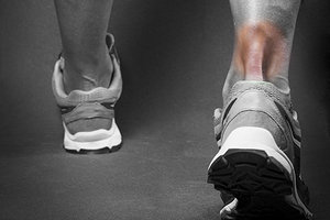
Achilles tendon problems are common in athletes, as well as among aging and active individuals. Those who run and/or climb stairs are particularly at risk of injury, but problems are also frequent in anyone who participates in activities such as racquet sports, volleyball and soccer.1
Functional Anatomy
The Achilles is the combined tendon of the gastrocnemius and soleus muscles. It is surrounded by a paratenon (rather than a synovial sheath), which is continuous with the fascia of the gastrocnemius and soleus muscles and the periosteum of the calcaneus. Tendinopathy usually occurs within the part of the tendon that is 2-6 cm proximal to the calcaneal insertion at a site of decreased vascularization.3
Poor blood flow could be a contributing factor to Achilles tendon overuse injuries, and especially tendon tears. Clement and his fellow authors speculate that "in individuals who overpronate, the conflicting internal and external rotatory forces imparted to the tibia by simultaneous pronation and knee extension may blanch or wring out vessels in the tendon and peritendon, causing vascular impairment and subsequent degenerative changes in the Achilles tendon."5
 This "region of relative avascularity" extends from 2 cm to 6 cm above the insertion into the calcaneus, and is the common site for a rupture of the Achilles tendon. This makes it especially important to ensure good blood flow as the body attempts to heal this condition.
Achilles Tendinopathy
The most common cause of functional breakdown of the Achilles tendon is a biomechanical deficit that places excess stress on the tendon. Inflexibilities, weakness and imbalances are noted throughout the lower extremity kinetic chain, into the pelvis and spine. In a study of 455 athletes with Achilles tendon injuries, Kvist discovered obvious biomechanical deficits in 60 percent of subjects.4 In many cases, the major biomechanical problem underlying an Achilles tendon problem is excessive pronation.
Most injuries of the Achilles tendon are "overuse" or "misuse" conditions caused by excessive and/or repetitive motion, often with poor biomechanics. The result is a microtrauma injury: The body is unable to keep up with the repair and re-strengthening needs, so the tissue begins to fail and becomes symptomatic. If it is not very painful (or when the pain is eliminated by pain-killing drugs), continued stress can lead eventually to complete failure, with a resulting acute tear of the tendon.
Because the Achilles tendon insertion on the calcaneus is medial to the axis of the subtalar joint, the calf muscles are powerful supinators of the subtalar joint. Therefore, when excessive pronation occurs, the Achilles tendon is overstressed, and eventually undergoes overuse degeneration and inflammation.
This was described by Clement and his fellow investigators. They explained how "pronation generates an obligatory internal tibial rotation, which tends to draw the Achilles tendon medially. Through slow motion, high-speed cinematography, we have seen that pronation produces a whipping action or bowstring effect in the Achilles tendon. This whipping action, when exaggerated, may contribute to microtears in the tendon, particularly in its medial aspect, and initiate an inflammatory response."5
These investigators believe that the control of functional overpronation with corrective orthotics is a necessary treatment for most patients with Achilles tendinitis.
Achilles Tendinitis / Tendinosis
It's not surprising that abnormal biomechanics of the foot and ankle can cause problems with the largest tendon in the leg. Symptoms are usually described as diffuse pain in or around the back of the ankle (from the calf to the heel). The pain is aggravated by activity, especially running uphill or climbing stairs, and relieved somewhat by wearing higher-heeled shoes or boots.
Palpation will reveal a tender thickening of the peritendon, and there could be crepitus during plantar and dorsiflexion. Often, patients report a recent increase in activity levels or a change in footwear. The most useful (and highly predictive) clinical test is to find a tender area of intratendinous swelling that moves with the tendon and disappears when the tendon is put under tension.6
Macroscopically, overused Achilles tendon tissues examined at surgery are dull, slightly brown and soft in comparison to normal tendon tissue, which is white, glistening and firm.7 There is a loss of collagen continuity and an increase in ground substance and cellularity, which is due to fibroblasts and myofibroblasts, and not inflammatory cells.8 This is why anti-inflammatory strategies (such as NSAIDS drugs and corticosteroid injections) are not indicated for these conditions, and actually could interfere with tendon repair.9
We now know that the condition we usually have described as "tendinitis" is actually better understood as "tendinosis," and is not due to inflammation, but an underlying degeneration of collagen tissues in response to mechanical overuse.10 In addition, patients with Achilles tendon problems must always be asked about their drug regimens – statins11 and fluoroquinolones (a class of antibiotic drugs)12 have been reported to cause Achilles tendinopathy and rupture.
Treatment Pearls
A combination of a change in activities and the use of custom-made functional orthotics has been found to be effective in most cases of Achilles tendinopathy.13 One of the most important treatment methods is to reduce any tendency to pronate excessively. In addition to custom-made orthotics, runners should wear well-designed shoes that provide good heel stability with a small amount of additional heel lift. Once any local inflammation has been controlled, treatments to improve the blood flow to the region of relative avascularity are necessary.
The use of corrective orthotics can prevent many overuse problems from developing in the lower extremities. As described above, most Achilles tendon problems develop from poor foot and ankle biomechanics, and control of pronation is needed to prevent recurrent injuries.14 Custom-made functional orthotics can support the hindfoot, midfoot and forefoot, providing biomechanical control throughout the entire gait cycle.
References
- Paavola M, Kannus P, Jarvinen TA, et al. Achilles tendinopathy. J Bone Joint Surg (U.S.), 2002;84:2026-76.
- Maffuli N, Kader D. Tendinopathy of tendo achillis. J Bone Joint Surg (U.K.), 2002;84:1-8.
- Carr AJ, Norris SH. The blood supply of the calcaneal tendon. J Bone Joint Surg (U.K.), 1989;71:100-01.
- Kvist M. Achilles tendon injuries in athletes. Ann Chir Gynaecol, 1991:80:188-201.
- Clement DB, et al. Achilles tendinitis and peritendinitis: etiology and treatment. Am J Sports Med, 1984;12:179-84.
- Maffuli N, Kenward MG, Testa V, et al. Clinical diagnosis of Achilles tendinopathy with tendinosis. Clin J Sports Med, 2003;13:11-5.
- Astrom M, Rausing A. Chronic Achilles tendinopathy: survey of surgical and histopathologic findings. Clin Orthop, 1995;316:151-64.
- Khan KM, Cook JL, Bonar F, Harcourt P, Astrom M. Histopathology of common tendinopathies: update and implications for clinical management. Sports Med, 1999;27:393-408.
- Almekinders LC, Temple JD. Etiology, diagnosis, and treatment of tendonitis: an analysis of the literature. Med Sci Sports Exerc, 1998;30:1183-90.
- Khan KM, et al. Overuse tendinosis, not tendinitis. Part 1: a new paradigm for a difficult clinical problem. Phys Sportsmed, 2000;28:38-48.
- Chazerain P, Hayem G, Hamza S, et al. Four cases of tendinopathy in patients on statin therapy. Joint Bone Spine, 2001;68:430-3.
- Vanek D, Sexena A, Boggs JM. Fluoroquinolone therapy and achilles tendon rupture. J Am Podiatr Med Assoc, 2003;93:333-5.
- Sorosky B, Press J, Plastaras C, Rittenberg J. The practical management of Achilles tendinopathy. Clin J Sport Med, 2004;14:40-4.
- Busseuil C, et al. Rearfoot-forefoot orientation and traumatic risk for runners. Foot & Ankle Intl, 1998;19:32-7.
Dr. John Hyland is an expert in the active rehabilitation of chiropractic patients. He was the developer and director of four chiropractic rehabilitation practices for more than eight years, and now consults, advises and trains DCs in the concepts and procedures of spinal rehabilitation.


