


Abdominal or "core" training is extremely popular. Traditional approaches have focused on sit-ups, while today, abdominal hollowing is the new trend. Core training should be as functional as possible and involve all the muscles of the core.
The Problem With the Sit-Up
A popular myth is that the sit-up is an ideal abdominal exercise. However, the sit-up places a high compressive load on the disc.18,19 It usually involves a posterior pelvic tilt, which unnecessarily elevates disc load.9 It commonly is performed early in the morning - a disaster waiting to happen, due to the morning's increased intradiscal pressure.2
Soloists vs. the Whole Orchestra
Spine stability is greatly enhanced by co-contraction (or co-activation) of antagonistic trunk muscles.6 Co-contractions increase spinal compressive load as much as 12 percent to 18 percent, or 440 Newtons, but they increase spinal stability even more, by 36 percent to 64 percent, or 2,925 Newtons.8 They have been shown to occur during most daily activities.16 This mechanism is present to such an extent that without co-contractions, the spinal column is unstable even in upright postures!22
 The lack of "core" muscle coordination has been found to be correlated to back pain. Researchers at Yale University have shown that a specific motor-control signature of delayed agonist-antagonistic muscle activation predicts which asymptomatic people will later develop low back pain (LBP).4 In particular, they noted longer muscle response latencies to perturbation in the "at risk" group than in healthy control subjects.
Inappropriate muscle activation patterns during seemingly trivial tasks (only 60 Newtons of force), such as bending over to pick up a pencil, can compromise spine stability and potentiate buckling of the passive ligamentous restraints.3 Certain times of the day, such as in the morning or after prolonged sitting, render the spine so unstable that if "surprised" by a trivial load, an injury can be precipitated.1
 Paul Hodges and colleagues from Australia have shown that delayed activation of the transverse abdominus muscle during arm or leg movements distinguishes between LBP patients and asymptomatic individuals.10,11 However, according to Canadian scientists, focusing on a single muscle is like focusing on a single guy wire.12 Research in Dr. Stuart McGill's laboratory at the University of Waterloo in Canada has found that the entire orchestra of muscles is responsible for spinal stability.12 The study demonstrated that different muscles play greater or lesser roles in the stability of the spine, depending on the activity/exercise, and concluded that no single muscle can be considered "the stabilizer of the spine."
Four Pillars in Training the Abdominal Wall
 Respiration: A common practice in gyms is to exhale with exertion. Unfortunately, many strenuous tasks occur when fatigue is setting in. If spine stability is compromised when one is gasping for air, the natural result will be low back injury (e.g., a deconditioned person shoveling snow). McGill demonstrated a loss of control of the "neutral spine posture" during weight-lifting under challenging aerobic circumstances.20 For all abdominal exercises, it is important that the patient is cued to maintain normal respiration.
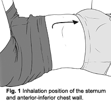
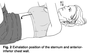
The diaphragm: A novel approach to achieving stronger co-activation of all abdominal wall muscles is to observe the position of the anterior chest wall.13 An elevated position is synchronized with the inhalation phase of respiration and will inhibit the normal postural function of the diaphragm. It is noted that the thoraco-lumbar (T/L) junction is hyperlordotic and the diaphragm is oblique in this position. (See Figure 1.) Ideally, a depressed (e.g., caudal) anterior chest position is facilitated, which is the "exhalation" position. In this case, the T/L junction is more neutral and the diaphragm is "centrated" in a horizontal position. (See Figure 2.) The "exhalation" position is believed to be facilitory of the abdominal wall, since active exhalation is produced by the abdominal muscles.
 Ask your patient to breathe in and feel how their chest naturally raises up. Then manually guide the chest down during exhalation and hold it there while the patient breathes in and out. Finally, ask your patient to actively hold their chest in this depressed exhalation position while continuing to breathe in and out.
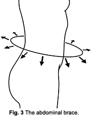
The abdominal brace: Co-contractions have been shown to occur automatically in response to unexpected or sudden loading and to have a stabilizing effect.14,17 Stokes22 has described how there basically are two mechanisms by which this co-activation occurs. One is a voluntary pre-contraction to stiffen the spinal column when faced with unexpected perturbations. The second is an involuntary, reflex contraction of the muscles quick enough to prevent instability following either expected or unexpected perturbations.7,14,17,22,23 Performing an abdominal brace (AB) is very simple. The patient should pretend they are about to be pushed or hit and they will "automatically" brace. (See Figure 3.)
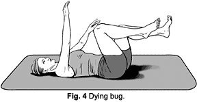
 Then the patient can be asked to hold the AB while breathing in and out. This takes practice. Finally, in recumbent, sitting, standing or various exercise positions (e.g., quadruped or "dying bug"), the AB can be maintained while the clinician offers slow and then quick perturbations in different planes. All the while, the patient is verbally cued to "breath and brace."
 Neutral spine posture: The fourth pillar for spine stability is maintenance of a "neutral spine" or normal lumbar lordosis. Many patients perform a posterior pelvic tilt, which actually places the lumbo-sacral spine in flexion and thus can potentially harm the disc via end-range loading in flexion. The "neutral zone is the inner region of a joint's range of motion (ROM), where minimal resistance to motion is encountered."21 According to McGill,19 "Because ligaments are not recruited when lordosis is preserved, nor is the disc bent, it appears that the annulus is at low risk for failure."
Some may wonder whether depressing the anterior rib cage to centrate the diaphragm will remove the lumbar lordosis. It can; however, it shouldn't since the key is to mobilize the rib cage inferiorly and posteriorly without altering the lumbo-sacral posture (e.g., lordosis). The mobilization should occur at the thoracolumbar junction while maintaining the lumbosacral junction in slight lordosis.
Training the Core
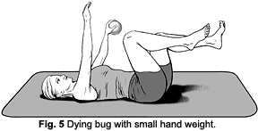
 The dying-bug position is ideal for core training. Begin in the recumbent squat position. This is the same position an infant can control at some time between 3 and 4 months old. The instruction is to tap a knee with the opposite hand and then partially separate the arm and leg, performing eight to 10 slow repetitions. (See Figure 4.) This can be progressed by adding a weighted medicine ball in the hand. (See Figure 5.)
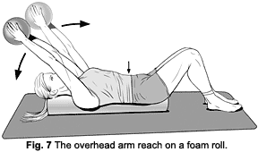
Another excellent recumbent core exercise is the overhead arm reach on a foam roll. (See Figure 7.) The same cueing is utilized to incorporate the four basic pillars of core training. Then have the patient slowly bring a medicine ball overhead without losing the exhalation position of the anterior chest. Move the ball back to the starting position and perform slow repetitions.
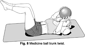
Since most functional activities require transverse plane motion (e.g., twisting), the trunk twist with a medicine ball is an ideal training exercise to re-educate the pattern of spine stability. (See Figure 8.) The key is to perform a small counter-rotation of only 1 or 2 inches. The arms and legs roll, but the spine should stay solidly in place on the mat.
 Abdominal exercises are utilized for cosmetic and therapeutic purposes. There are a number of myths which should be unmasked about this subject regarding sit-ups, morning exercise, the posterior pelvic tilt, the transverse abdominus, exhaling with exertion, etc. The four pillars of core training described in this article should be incorporated into all abdominal training. The dying bug is an excellent starting point to "groove" healthy motor patterns. This should be seen as a first step, but not an end in itself. Progressions to side support, quadruped, and most importantly, upright (e.g., squat, lunge, push, and pull) positions are essential to ensure sufficient stability in the lumbar spine during participation in job demands, daily activities, as well as sports and recreational activities.
References
- Adams MA, Dolan P. Recent advances in lumbar spine mechanics and their clinical significance. Clin Biomech, 1995;10:3-19.
- Adams MA, Hutton WC. Gradual disc prolapse. Spine, 1985;10:524-31.
- Andersson GBJ, Winters JM. Role of muscle in postural tasks: spinal loading and postural stability. In: Multiple Muscle Systems, Winters JM, Woo SL-Y, Eds. New York: Springer-Verlag; 23:375-95.
- Cholewicki J, Silfies SP, Shah RA, et al. Delayed trunk muscle reflex responses increase the risk of low back injuries. Spine, 2005;30(23):2614-20.
- Cholewicki J, Panjabi MM, Khachatryan A. Stabilizing function of the trunk flexor-extensor muscles around a neutral spine posture. Spine, 1997;22:2207-12.
- Cholewicki J, McGill SM. Mechanical stability of the in vivo lumbar spine: Implications for injury and chronic low back pain. Clin Biomech, 1996;11(1):1-15.
- Cresswell AG, Oddsson L, Thorstensson A. The influence of sudden perturbations on trunk muscle activity and intra-abdominal pressure while standing. Exp Brain Res, 1994;98:336-41.
- Granata KP, Marras WS. Cost-benefit of muscle cocontraction in protecting against spinal instability. Spine, 2000;25:1398-1404.
- Hickey DS, Hukins DWL. Relation between the structure of the annulus fibrosis and the function and failure of the intervertebral disc. Spine, 1980;5:106-16.
- Hodges PW, Richardson CA. Delayed postural contraction of the transverse abdominus associated with movement of the lower limb in people with low back pain. J Spinal Disord, 1998;11:46-56.
- Hodges PW, Richardson CA. Altered trunk muscle recruitment in people with low back pain with upper limb movements at different speeds. Arch Phys Med Rehabil, 1999;80:1005-12.
- Kavcic N. Grenier S, McGill SM. Determining the stabilizing role of individual torso muscles during rehabilitation exercises. Spine, 2004;29:1254-65.
- Kolar P. Facilitation of Agonist-Antagonist Co-Activation by Reflex Stimulation Methods. In: Rehabilitation of the Spine: A Practitioner's Manual, 2nd ed., Liebenson C, Ed. Philadelphia: Lippincott, Williams & Wilkins, 2007.
- Lavender SA, Mirka GA, Schoenmarklin RW, et al. The effects of preview and task symmetry on trunk muscle response to sudden loading. Human Factors, 1989;31:101-15.
- Liebenson C. Functional Stability Training. In: Rehabilitation of the Spine: A Practitioner's Manual, 2nd ed., Liebenson C, Ed. Philadelphia: Lippincott, Williams & Wilkins, 2007.
- Marras WS, Mirka GA. Muscle activities during asymmetric trunk angular accelerations. J Orthop Res, 1990;8:824-32.
- Marras WS, Rangarajulu SL, Lavender SA. Trunk loading and expectation. Ergonomics, 1987;30:551-62.
- McGill SM. Lumbar Spine Stability: Mechanism of Injury and Restabilization. In: Rehabilitation of the Spine: A Practitioner's Manual, 2nd ed., Liebenson C, Ed. Philadelphia: Lippincott, Williams & Wilkins, 2007.
- McGill SM. Ultimate Back Fitness and Performance, 2nd ed. Waterloo, ON, Canada: Wabunu, 2006.
- McGill SM, Sharratt MT, Seguin JP. Loads on the spinal tissues during simultaneous lifting and ventilatory challenge. Ergonomics, 1995;38:1772-92.
- Panjabi MM. The stabilizing system of the spine. Part 1. Function, dysfunction, adaptation, and enhancement. J Spinal Disorders, 1992;5:383-9.
- Stokes IAF, Gardner-Morse M, Henry SM, Badger GJ. Decrease in trunk muscular response to perturbation with preactivation of lumbar spinal musculature. Spine, 2000;25:1957-1964.
- Wilder DG, Aleksiev AR, Magnusson ML, et al. Muscular response to sudden load. a tool to evaluate fatigue and rehabilitation. Spine, 1996;21:2628-39.
Click here for previous articles by Craig Liebenson, DC.