


A negative Derifield (-D) occurs when the reactive (shorter) leg in the prone position becomes shorter, stays short, or fails to cross over in the flexed position.1-2 The negative Derifield test result suggests a θY-axis segmental dysfunction manifesting as rotation of the ilium or sacrum relative to the longitudinal axis of the body.
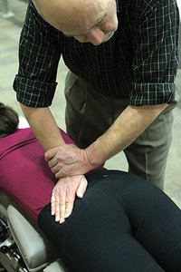 Fig. 1: Left SI fluid motion, performed to determine sacroiliac fixation.
Four different listings of sacroiliac joint dysfunction may be suggested by a negative Derifield test. We have already addressed the external (EX) ilium or posterior rotation of the sacral base on the left or right (P-L or P-R.) We will now address the other possibilities: internal (IN) ilium or anterior rotation of the sacral base on the left or right (A-L or A-R.)
Fig. 1: Left SI fluid motion, performed to determine sacroiliac fixation.
Four different listings of sacroiliac joint dysfunction may be suggested by a negative Derifield test. We have already addressed the external (EX) ilium or posterior rotation of the sacral base on the left or right (P-L or P-R.) We will now address the other possibilities: internal (IN) ilium or anterior rotation of the sacral base on the left or right (A-L or A-R.)
Determining the Listing
Use the θY-axis segmental dysfunction test5 to determine the listing by lifting the leg on the side of fixation into extension [as discussed in part 2 – May 15 issue]. If the reactive leg stays short in the prone extended position, flex the knees and observe for changes in leg length on the side tested. If the leg becomes shorter in the flexed position, the test suggests internal (IN) ilium or anterior rotation of the sacrum on the side tested. IN ilium tends to predominate.
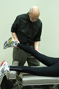 Fig. 2: Left θY-axis segmental dysfunction test for the pelvis. This is an isolated test performed immediately following a -D finding to narrow the possible θY-axis pelvic misalignments from four to two possibilities.
Determine the appropriate listing and line of correction using an articular pressure test.3-5 To test for an IN ilium, stand on the side opposite involvement at the level of the SI joint. Contact the medial aspect of the PSIS, and apply an arcing tissue pull from superior to inferior and medial to lateral. If the legs become even in the extended and flexed position, the test is positive for an IN ilium. (Recall that an IN ilium tends to ride up the articulation.)
Fig. 2: Left θY-axis segmental dysfunction test for the pelvis. This is an isolated test performed immediately following a -D finding to narrow the possible θY-axis pelvic misalignments from four to two possibilities.
Determine the appropriate listing and line of correction using an articular pressure test.3-5 To test for an IN ilium, stand on the side opposite involvement at the level of the SI joint. Contact the medial aspect of the PSIS, and apply an arcing tissue pull from superior to inferior and medial to lateral. If the legs become even in the extended and flexed position, the test is positive for an IN ilium. (Recall that an IN ilium tends to ride up the articulation.)
Adjusting an IN Ilium
To adjust an IN ilium, stand on the side opposite the involved joint, contact the medial aspect of the PSIS, and apply a thrust posterior to anterior, medial to lateral, and superior to inferior through the plane line of the joint space. (Note: The line of correction for the thrust is identical to the direction of the articular pressure test.) Following the adjustment, legs should be even in both the extended and flexed positions.1-3 Evaluate for mobility with the SI fluid motion test [as discussed in part 1 – March 1 issue]. The joint will now typically be freely movable.
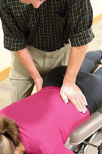 Fig. 3: Adjusting setup for left anterior rotation of the sacrum (A-R).
If the articular pressure test for the IN ilium does not cause the legs to become even in the extended and flexed position, test for the last option, anterior rotation of the sacrum (A-L or A-R).1-2 Take a scissors stance on the side of involvement at the level of the distal femur. With superior hand help palm-up, slide the fingers from lateral to medial approximately 2 inches bellow the anterior superior iliac spine to contact the mid-lateral aspect of the inguinal ligament. (Recall that the inguinal ligament runs medial and inferior from the ASIS to the pubic tubercle.) Use finger pads 2-4 to apply a straight lateral-to-medial pressure against the mid-lateral aspect of the inguinal ligament. If the legs become even in the extended and flexed position, the test is positive for anterior rotation of the sacrum on the side tested.
Fig. 3: Adjusting setup for left anterior rotation of the sacrum (A-R).
If the articular pressure test for the IN ilium does not cause the legs to become even in the extended and flexed position, test for the last option, anterior rotation of the sacrum (A-L or A-R).1-2 Take a scissors stance on the side of involvement at the level of the distal femur. With superior hand help palm-up, slide the fingers from lateral to medial approximately 2 inches bellow the anterior superior iliac spine to contact the mid-lateral aspect of the inguinal ligament. (Recall that the inguinal ligament runs medial and inferior from the ASIS to the pubic tubercle.) Use finger pads 2-4 to apply a straight lateral-to-medial pressure against the mid-lateral aspect of the inguinal ligament. If the legs become even in the extended and flexed position, the test is positive for anterior rotation of the sacrum on the side tested.
Adjusting for Anterior Rotation of the Sacrum
To adjust for anterior rotation of the sacrum, raise the table and reposition the patient in the supine position. (Remember, the side you just tested is now on the opposite side of the table.) Stand on the side of involvement in a shortened scissors stance and use the superior (away) hand for the contact. Have the patient flex the opposite hip and knee, and place the heel at the bottom of the table. Use the inferior (near) hand to stabilize the thrust by placing the palm over the distal femur, just above the knee.
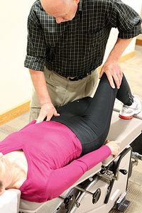 Fig. 4: Adjusting setup for left IN ilium.
Using the superior hand, position the finger web between the second and third fingers against the inferior aspect of the ASIS. Contact the mid-lateral aspect of the inguinal ligament with the thenar at the base of the thumb, taking a lateral-to-medial tissue pull. Apply an arcing thrust to the ligament lateral to medial and inferior to superior by torquing the elbow in toward the patient.
Fig. 4: Adjusting setup for left IN ilium.
Using the superior hand, position the finger web between the second and third fingers against the inferior aspect of the ASIS. Contact the mid-lateral aspect of the inguinal ligament with the thenar at the base of the thumb, taking a lateral-to-medial tissue pull. Apply an arcing thrust to the ligament lateral to medial and inferior to superior by torquing the elbow in toward the patient.
The adjustment works best with a pelvic drop piece set to minimal tension. Following the adjustment, legs should be even in both the extended and flexed positions. Finally, evaluate for mobility with the SI fluid motion test. The joint will now typically be freely movable.
The six primary misalignments of the sacroiliac joints discussed in this series highlight the use of the positive and negative Derifield leg check to guide clinical decision-making. Additional misalignments are possible, but relatively rare. Therefore, the strategies presented will resolve most presenting complaints of sacroiliac dysfunction.
References
- Laufenberg PC. Thompson Terminal Point Handbook. Davenport, IA: J Clay Thompson, 1974.
- Thompson Educational Workshops. The Thompson Technique Reference Manual. Elgin, IL: 1984.
- Fuhr AW, et al. Activator Methods Chiropractic Technique, 2nd Edition. St. Louis: Mosby-Elsevier, 2009.
- Haas M, Peterson D, Panzer D, et al. Reactivity of leg alignment to articular pressure testing: evaluation of a diagnostic test using a randomized crossover clinical trial approach. J Manip Physiol Ther, 1993;16(4):220-7.
- Khauv KB, John C. Health-related quality of the improvements in adult patients with chronic low back pain under low-force chiropractic care: a practiced-based study. Chiropr J Aust, 2011 Dec;41(4):118-122.
Dr. Howard Pettersson, a 1976 graduate of Logan College of Chiropractic, is an associate professor of technique at Palmer College of Chiropractic. He was the senior editor of Activator Methods Chiropractic Technique – College Edition, published in 1989, and published Pelvic Drop Table Adjusting Technique in 1999. His most recent publication, written with Dr. Green, is How to Find a Subluxation, published in 2003.
Dr. J.R. Green is a 1988 Graduate of Palmer College of Chiropractic. He retired from the Palmer faculty after many years of teaching basic sciences and chiropractic technique. He is currently in private practice in Galva, Ill., and is also an adjunct professor of chemistry with the Eastern Iowa Community College District. Dr. Green was one of the writers of Activator Methods Chiropractic Technique (1997) and also worked as a technical writing consultant on Activator Methods Chiropractic Technique – College Edition and Pelvic Drop Table Adjusting Technique.