According to Dr. Voyer, the local effects of this stretch are zygopophyseal separation, imbibition of the disc, increased venous return, normalization of muscle tone (by extreme eccentric contraction), proprioceptive facilitation of the paraspinal muscles, and improved kinetic sense. General effects from this procedure include normalization of myofascial tensions, decreased psychomotor barriers, and an increased well-being. Putting the myofascial chains around a primary lesion (the local spinal decoaptation) into tension will result in an increase in postural normalization. Patients report that after this type of stretch, not only do their back pains decrease, but other symptoms also improve.
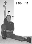
The author demonstrates the T10-T11 stretch.
The accompanying photo shows the position for a specific stretch for the T10-T11 disc level.
These stretches are not easy to perform. Several visits are required to make sure the patient is in the correct posture. For some spinal segments, the patient cannot create the total stretch at first, but over time it may be accomplished. Inability to perform the stretch is sometimes valuable in finding where other fascial/structural problems may be located.
The right hip is flexed straight back as far as it can go. The right foot is resting on the heel in dorsiflexion and eversion. The left lower extremity is stretched out at 45 degrees with the foot dorsiflexed and inverted. The shoulders and spine are pressing against the wall. The right arm is fully extended vertically with the wrist dorsiflexed, and the arm and hand externally rotated. The chin is retracted and the head is extended vertically. All stretching and extending is done to the maximum and held for a minute. In this position, if everything is correct, the patient will feel the tension specifically at the T10-T11 level. Good luck.
Warren Hammer,MS,DC,DABCO
Norwalk, Connecticut
Click here for previous articles by Warren Hammer, MS, DC, DABCO.


