Surprisingly, the motor control system functions well when under load. Muscles stabilize joints by stiffening like rigging on a ship (McGill, 2000), but when load is minimum, such as when the body is relaxed or a task is trivial, the motor control system is often "caught off guard" and injuries are often precipitated. According to Bogduk and Twomney, "After prolonged strain, ligaments, capsules and IV discs of the lumbar spine may creep, and they may be liable to injury if sudden forces are unexpectedly applied during the vulnerable recovery phase." (Bogduk, Twomney 1991)
Most low back injuries are not the result of a single exposure to a high- magnitude load, but instead a cumulative trauma from sub-failure magnitude loads: for instance, repeated small loads (e.g., bending) or a sustained load (e.g., sitting). In particular, low back injury has been shown to result from repetitive motion at end range. According to McGill, it is usually a result of "a history of excessive loading which gradually but progressively reduces the tissue failure tolerance." (McGill, 1998)
Epidemiologic studies on gymnasts and Australian cricket bowlers demonstrates that posterior arch damage leading to spondylolysthesis is not just due to shear forces, but is also correlated with repetitive full-range activities (Hardcastle et al., 1992). Disc injury has been shown to be related to three factors: full end-range flexion in younger spines (due to higher water content) (Adams, Hutton 1982, Adams, Hutton 1985; Adams, Muir 1976); repetitive end-range flexion loading motion in excess of 20-30,000 cycles (King, 1993; Gordon et al., 1991); and, an epidemiological association between disc herniation and sedentary sitting occupations (Videman et al., 1990).
The back is more vulnerable at certain times of the day. In the first hour after awakening, or after prolonged static full flexion such as sitting or stooping, the body is at greatest risk. Disc bending stresses are increased by 300% and ligaments by 80% in the morning (Adams et al., 1987). McGill has reported that after just three minutes of full flexion, subjects lost half their stiffness (McGill 1999).
How Does the Body Resist Injury?
Support and stability to the low back arise from muscles. Certain muscles in particular have been shown to stabilize the low back in various situations. Surprisingly, the one muscle that is highly active during flexion, extension and lateral bending tasks is the quadratus lumborum (McGill 1996). Its architecture is ideally suited to be a stabilizer, since it attaches each transverse process to the more rigid pelvis and rib cage, facilitating a bilateral buttressing effect for the vertebrae (McGill 2000).
Interestingly, it is not strength but coordination between agonist and synergist muscles that plays a pivotal role in resisting injury. Sparto et al. reported spinal loading forces were increased during a fatiguing isometric trunk extension effort without a loss of torque output (Sparto 1997). Torque output remained constant because as the erector spinae fatigued, substitution by secondary extensors such as the internal oblique and latissmus dorsi muscles occurred.
Agonist-antagonist muscle co-activation is an important mechanism of how joint injury is prevented. Cholewicki et al. examined the theory that antagonistic trunk muscle coactivation is necesssary to provide mechanical stability to the lumbar spine around a neutral posture (Cholewicki 1997). The authors found that antagonistic muscle coactivation increased in response to increased axial load on the spine.
According to Panjabi, the following subsystems work together to promote stability (Panjabi 1992):
- central nervous subsystem (control)
- osteoligamentous substystem (passive)
- muscle subsystem (active)
Panjabi explains that "The neural subsystem receives information from the transducers, determines specific requirements for spinal stability, and causes the active subsystem to achieve the stability goal." (Panjabi 1992). Good motor control and appropriate activities maintain movements in the neutral or elastic range. Poor motor control or inappropriate activities allow movement to reach the elastic range.
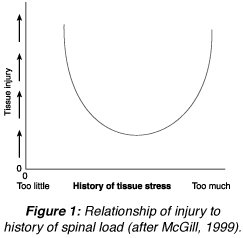
Figure 1: Relationship of injury to history of spinal load (after McGill, 1999).
Injury Prevention
To avoid injury, conditioning or adaptation must keep pace as exposure to external load increases. Exposure to load must be removed so that the normal healing/adaptation process can fulfill its objective of increasing the failure tolerance of the tissues. Too little or too infrequent exposure to external load and conditioning never raises tissue failure tolerance. Too much, too frequent or too prolonged exposure, and adaptation can't keep pace. McGill has shown this relationship between tissue loading and risk of injury as "U" shaped (McGill 2000).
How can knowledge of the mechanism of injury influence our management? According to McGill, "Evidence from tissue-specific injury generally supports the notion of a neutral spine (neutral lordosis) when performing loading tasks to minimize the risk of low back injury ... Avoiding spine end range of motion during activity can reduce the risk of several types of injury." (McGill 1998)
Knowledge of when injury is most likely to occur can also influence what we do. The early morning or after prolonged sitting are particularly vulnerable times. Reilly et al. (Reilly et al., 1984) showed that 54% of the loss of disc height (water content) occurs in the first 30 minutes after arising. Disc bending stresses are increased significantly in the morning (Adams et al., 1987). After just a brief period of sitting or stooping in end range, protective joint stiffness is compromised (McGill 1999). Even after 30 minutes of rest, residual joint laxity persisted! Therefore, avoidance of high-risk activities early in the morning or after sitting or stooping in full flexion is crucial to injury or reinjury prevention.
Teaching workers to lift with their knees, not their backs, is overly simplistic. Most workers have learned various techniques to avoid injury that are inconsistent with this advice. Better advice is consistent with the following principles: avoid end-range motion; rotate jobs to vary loads; allow frequent rest breaks; and keep loads close to the spine (McGill, Norman 1993).
Axler and McGill have demonstrated that both muscle output and spinal load can be measured for a variety of exercises (Axler, McGill 1997). Muscle output is determined as a percentage of maximum voluntary contraction ability (MVC) and spinal load as a measure of spinal compression and shear forces. Ideal exercises have a high ratio of muscle challenge to spinal load. Such analyses give surprising data about common exercises prescribed for low back pain. For instance, spinal load is not different during sit-ups with knees bent or straight. In either case, the load is over 3000N and should not be prescribed in the low back recovering population! (Axler, McGill 1997; McGill 1995). There are safer back exercises.
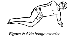
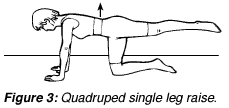
Safe back principles have emerged from rigorous analysis of biomechanical and kinesiological aspects of spinal function (McGill 1998b; Liebenson 1999). By emphasizing endurance training of key spinal stabilizers, positive outcomes have been achieved (Hides et al., 1996; Timm et al., 1994; O'Sullivan et al., 1997). These principles can be summarized into a user-friendly approach which will enhance clinical application and patient compliance.
Figure 2: Side bridge exercise.
Figure 3: Quadruped single leg raise.
The main emphasis of rehabilitative exercise is to train "inner range" endurance. This requires kinesthetic awareness of "neutral spine" postures and regular sustained training of this motor control against a variety of challenges. Multiple reps of 5-6 second holds daily should be performed. According to Manniche et al., up to three months may be required to achieve a long-lasting beneficial effect (Manniche et al., 1991).
References
- Adams MA, Hutton WC. Prolapsed intervertebral disc: a hyperflexion injury. Spine 1982;7:184. Adams MA, Hutton WC. Gradual disc prolapse. Spine 1985;10:524.
- Adams MA, Dolan P, Hutton WC. Diurnal variations in the stresses on the lumbar spine. Spine 1987;12(2);130.
- Adams P, Muir H. Qualitative changes with age of proteoglycans of human lumbar discs. Ann Rheum Dis 1976;35:289.
- Axler CT, McGill SM. Low back loads over a variety of abdominal exercises: searching for the safest abdominal challenge. Med Sci Sports Exerc 1987;29:804-810.
- Bogduk N, Twomney L. Clinical Anatomy of the Lumbar Spine, 2nd edition. Churchill Livingstone, 1991.
- Cholewicki J, Panjabi MM, Khachatryan A. Stabilizing function of the trunk flexor-extensor muscles around a neutral spine posture. Spine 1997;19:2207-2212.
Gordon SJ, et al. Mechanism of disc rupture: a preliminary report. Spine 1991;16:450. - Hardcastle P, Annear P, Foster D. Spinal abnormalities in young fast bowlers. J Bone Jt Surg 1992;74B(3):421.
- Hides JA, Richardson CA, Jull GA. Multifidus muscle recovery is not automatic after resolution of acute, first-episode of low back pain. Spine 1996;21(23):2763-2769.
- King AI. Injury to the thoracolumbar spine and pelvis. In: Nahum AM, Melvin JW (eds). Accidental Injury, Biomechanics and Presentation. New York: Springer-Verlag, 1993. Liebenson CS. The safe back workout. JNMS 1999;7(1).
- Manniche C, Lundberg E, et al. Intensive dynamic back exercises for chronic low back pain. Pain 1991;47:53-63.
- McGill SM, Norman RW. Low back biomechanics in industry: the prevention of injury through safer lifting. In: Grabiner M (ed.) Current Issues in Biomechanics. Champaign, IL: Human Kinetics, 1993.
- McGill S. The mechanics of torso flexion: sit-ups and standing dynamic flexion maneuvers. Clin Biomech 1995;10:184-192.
- McGill SM, Juker D, Kropf P. Quantitative intramuscular myolectric activity of the quadratus lumborum during a wide variety of tasks. Clin Biomechanics 1996;11(3):170-2.
- McGill SM. Low back exercises: prescription for the healthy back and when recovering from injury. In: Resources Manual for Guidelines for Exercise Testing and Prescription, 3rd ed. Indianapolis, IN: American College of Sports Medicine; Baltimore, Williams and Wilkins, 1998.
- McGill SM. Low back exercise: evidence for improving exercise regimens. Phys Ther 1998b;78:754-765. McGill SM. Lecture at LACC, September 18, 1999.
- McGill SM. Clinical biomechanics of the thoracolumbar spine. In: Dvir Z (ed.) Clinical Biomechanics, in press (sched pub. 2000).
- O'Sullivan P, Twomey L, Allison G. Evaluation of specific stabilizing exercise in the treatment of chronic low back pain with radiologic diagnosis of spondylolysis or spondylolysthesis. Spine 1997;24:2959-2967.
- Panjabi MM. The stabilizing system of the spine. Part 1. Function, dysfunction, adaptation and enhancement. J Spinal Disorders 1992;5:383-389.
- Reilly T, Tynell A, Troup JDG. Circadian variation in the human stature. Chronobiology It 1984;1:121.
- Sparto PJ, Paarnianpour M, Massa WS, Granata KP, Reinsel TE, Simon S. Neuromuscular trunk performance and spinal loading during a fatiguing isometric trunk extension with varying torque requirements. Spine 1997;10:145-156.
- Timm KE. A randomized control study of active and passive treatments for chronic low back pain following L5 laminectomy. JOSPT 1994;20:276-286.
- Videman T, Nurminen M, Troup JDG. Lumbar spinal pathology in cadaveric material in relation to history of back pain, occupation and physical loading. Spine 1990;15(8):728.
Click here for previous articles by Craig Liebenson, DC.


