In our first article in this series, we suggested that effective care of recurrent sacroiliac joint (SIJ) and related low back pain should include, in addition to osseous adjusting, attention to musculature and other soft tissue affecting SIJ function. Specifically, hamstrings, gluteal muscles and the iliopsoas complex are critical in stabilizing the SIJ with regard to force closure.
PI Ilium Misalignment
Let us first address one of the more common osseous misalignments of the SIJ, the posterior inferior (PI) ilium. The osseous component may be described as an ilium that is displaced posteriorly and inferiorly relative to sacrum. The posterior superior iliac spine (PSIS) on the side of involvement may palpate relatively inferior to the non-involved side. The biomechanical issue in such cases is a dysfunction of form closure, as described by Vleeming.1
In her discussion of anterior pelvic crossed syndrome, Key2 describes what chiropractors have traditionally referred to as "posterior pelvic tilt." The two major components of this finding are decreased sacral base angle (less than 33 degrees on radiographic analysis) and hypolordosis of the lumbar spine. The patient often presents with a characteristic "flat back and tucked butt" gait and standing posture. Our hypothesis is that PI ilium is the more common misalignment of the SIJ.
Assessment of the PI Ilium
To assess a PI ilium, the following observations and tests may yield the optimum clinical results:
Side-Lying Iliac Compression – The patient lies on the table with the symptomatic side up. Use the heel of the hand just lateral and anterior to the PSIS to apply a firm lateral to medial pressure through the SIJ. Provocation of pain suggests involvement of the SIJ.
Lewin-Gaenslen – The patient is in the side-lying position, involved side up. The examiner passively extends the upper leg to the limit of extension at the hip and SIJ, while the patient pulls the lower leg into acute flexion of the hip. Provocation of pain suggests SIJ involvement.
Flexed Thigh Thrust – The patient is supine and the examiner stands on the asymptomatic side. Flex the thigh on the side of involvement to 90 degrees and position the inferior hand between the patient's sacrum and the table to block the sacrum. With the superior hand, apply a thrust through the longitudinal axis of the femur to create a shearing stress to the SIJ. Reproduction of pain suggests SIJ involvement.
Anterior Distraction – The patient is supine with a towel or padding under the low back to maintain the lumbar curve. The examiner takes a stance perpendicular to the patient at the mid-thigh level. Cross the forearms, contact the anterior superior iliac spines with heels of both hand, and apply a steady posterior pressure through the SIJs. Provocation of pain suggests SIJ involvement.
Prone Sacral Thrust – The patient is prone and the examiner takes a straight-on stance to the patient's side. Place the heel of the inferior hand at the level of the third sacral tubercle and use the superior hand to stabilize the contact hand. Apply three to five thrusts in a posterior-to-anterior direction through the body of the sacrum. Reproduction of pain suggests SIJ involvement.
Consensus supports a clinical impression of SIJ abnormality if any two of the above tests are positive.3
Discussion
Evidence of an uncomplicated PI ilium includes observation of leg-length inequality with the apparent shorter leg on the side of involvement. A relative toe-out foot flare tends to occur on the side of the PI ilium and the foot on the involved side typically shows relative inversion.4 A positive sacroiliac fluid motion test indicates fixation of the SIJ on the side of involvement. Usually the observations support proceeding with osseous adjusting.
When osseous adjustment of the PI ilium does not yield satisfactory outcomes, it is time to look at complicating factors such as contracture of hamstring and an iliopsoas muscles, among others, myofascial distortions of the pelvic, lumbar, and hip soft tissues, or disruptions of the kinematic chain involved with posture and locomotion. Unsatisfactory results of osseous adjustment may include persistence or recurrence of pain, distortions of posture, stance, and gait, and interference with activities of daily living.
Soft-tissue complications of SIJ fixation may be of acute onset, but are more often gradual and chronic. Consequently, chiropractic care may require time and persistence to be successful. Furthermore, we concur with Heller5 that an otherwise healthy SIJ is inherently stable (form closure), but we suggest stressors to periarticular soft tissue – muscles, tendons, ligaments and fascia – can destabilize the function of the SIJ (force closure).
SI Fluid Motion Test
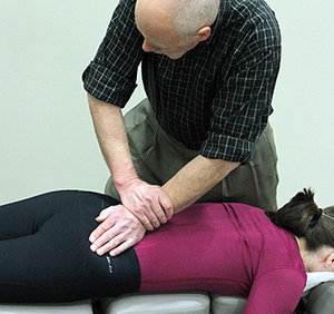
 FIG 1 Right SI Fluid Motion Test
To begin, test the patient for sacroiliac joint fixation using the sacroiliac fluid motion test. To perform the test, apply a firm, continuous pressure to the medial, superior aspect of the PSIS anterior, inferior and lateral toward the acetabulum. While performing the test, observe the leg-length changes in response to applying fluid motion through the sacroiliac joint space.
If only the leg on the side tested elongates, the joint is not fixed. If, on the other hand, both legs elongate simultaneously during the test, the joint is fixed. If fixation is found on the side of the PI foot, then adjust for PI ilium.
PI Ilium Adjustment
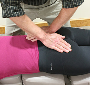
 FIG 2 Right PI Llium Adjustment
Utilize dual segmental contact points. Place the inferior (contact) hand fleshy pisiform on the inferior aspect of the PSIS. Place the superior (contact) hand thenar on the involved side sacral ala at the level of S2-S3. Deliver three toggle-like thrusts. Thrust equally and simultaneously with both hands.
Thrust inferior to superior (I-S) and posterior to anterior (P-A) with the inferior hand, and superior to inferior (S-I) and posterior to anterior (P-A) with the superior hand. Avoid following the thrust beyond the excursion of the drop mechanism and release quickly.
Clinical Pearls
The functionally unstable SIJ typically responds to a combination of osseous adjusting and soft-tissue treatment. Since the hamstrings and piriformis are facilitated and shortened, and the iliopsoas and glutei muscles are inhibited and lengthened with an anterior pelvic crossed syndrome (posterior pelvic tilt), a chronic PI ilium misalignment is often observed. Therefore, a therapeutic approach should also include passive stretching and soft-tissue manipulation of those muscles. Furthermore, teaching the patient to work those muscle by stretching and by correcting standing and sitting posture will be a positive step in stabilizing the sacroiliac joint, improving mobility, and minimizing pain and discomfort.
References
- Vleeming A, Schuenke M. Form and force closure of the sacroiliac joints. Phys Med & Rehabil, 2019 Aug;11 Suppl 1:S24-S31.
- Key J, et al. A model of movement dysfunction provides a classification system guiding diagnosis and therapeutic care in spinal pain and related musculoskeletal syndromes: a paradigm shift - part 2. J Bodywork Movement Ther, 2008;12:105-20.
- McGarth MC. Composite sacroiliac joint pain provocation tests: a question of clinical significance. Int J Osteopathic Med, 2010;13:24-30.
- Cooperstein R. "The Reverse Double Whammy Leg Check." Dynamic Chiropractic, July 28, 1997.
- Heller M. "SI Stability - or Something Else?" Dynamic Chiropractic, Dec. 1, 2019.
Dr. Howard Pettersson, a 1976 graduate of Logan College of Chiropractic, is an associate professor of technique at Palmer College of Chiropractic. He was the senior editor of Activator Methods Chiropractic Technique – College Edition, published in 1989, and published Pelvic Drop Table Adjusting Technique in 1999. His most recent publication, written with Dr. Green, is How to Find a Subluxation, published in 2003.
Dr. J.R. Green is a 1988 Graduate of Palmer College of Chiropractic. He retired from the Palmer faculty after many years of teaching basic sciences and chiropractic technique. He is currently in private practice in Galva, Ill., and is also an adjunct professor of chemistry with the Eastern Iowa Community College District. Dr. Green was one of the writers of Activator Methods Chiropractic Technique (1997) and also worked as a technical writing consultant on Activator Methods Chiropractic Technique – College Edition and Pelvic Drop Table Adjusting Technique.
Dr. D. Ranier Pavilek graduated as a certified athletic trainer from BYU in 1991, and from Palmer College in 2001. He is currently a faculty clinician in the Chiropractic Rehabilitation and Sports Injury Department, Palmer Chiropractic Clinics. He also teaches Care and Prevention of Athletic Injuries and Kinesiology courses in Palmer's undergraduate program and developed the Advanced Soft Tissue course in Palmer's elective program.


