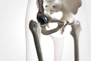
A joint replacement is expected to last 15-20 years; maybe longer with the newer devices, but the population is living longer and more people expect to have a joint replacement as a matter of medical management for their aging joints.
More than 7 million people in the U.S. are living with a least one joint replacement.1 Because our patient population tends to be older, one could assume the prevalence of joint replacements would be higher in chiropractic patients. My point is that we will be treating more patients with joint replacements and should be aware of the additional complications associated with this surgery. We need to be able to identify patients who may be at risk for prosthetic instability.
Prosthetic Instability: Spot the Signs, Understand the Causes
The prosthetic (artificial) components are either cemented (attached with bone cement) or hammered into the bone so they will eventually attach to patient's bone. If the bone fails to grow onto the components or the cement loosens over time, the joint will feel painful, unstable or loose. Excess body weight, along with high-impact activities or exercise, puts stress on the joint components.
Of course, there is also just the wear and tear of the components over time. Most initial joint replacements are designed to last 15-20 years. The younger and more active the patient, the more likely there will be a need for a revision surgery, as the components naturally will wear out over time.
 The signs of joint replacement failure are: loosening or instability, infection, frequent or recurring hip dislocations, fracture, and metal allergy. Loosening or instability is the most common sign in older joint replacements.
The most common symptoms associated with loosening or instability of a joint replacement include: pain; popping or clicking; a sensation that the joint is moving in the socket; hip subluxation; full hip dislocation; and a sensation that the joint is "giving out" when weight-bearing.
Infection occurs more often shortly after surgery, but also can occur years later. Pain and swelling are the usual initial clinical presentations. Metal allergy is more difficult to diagnose because it presents similarly to infection.
If the hip replacement was done prior to May 2016, the patient may have received a metal-on-metal device. The wear and tear on these metal components release microscopic metal particles. This may lead to sensitivity or an allergic reaction in some patients. (Metal-on-metal hip replacements are no longer used in the U.S.)
If the patient received a hip replacement before 2016, the type of joint replacement should be documented in their medical record. If the patient presents with any clinical symptoms suggesting infection or instability, they need to be referred to a specialist.
Joint Replacement Dislocation
Dislocation is the second most common complication following THA. Most dislocations occur early in the post-operative period; usually during the first 12 weeks. Causes for early dislocation include patient-associated factors and surgical factors. Two-thirds of the time, these cases can be treated conservatively with reduction; however, one-third will require surgical treatment.
These early dislocations we are not likely to see. The later dislocations, which are due primarily to instability caused by loosening of the components and polyethylene-metallic wear and tear, we may see in our clinic practice.
The long-term threat with hip replacements is the interaction between surfaces of the components, causing debris,, which can cause inflammation in adjacent tissues that result in bone loss; in turn leading to prosthesis loosening. Of course, there are other contributing factors including fracture, malpositioning of components, infection around the implant, poor cementing technique, and certain stem designs prone to loosening.3
How You Can Help Prolong Joint Replacement Longevity
The goal is to make the joint replacement last as long as possible; which means avoiding issues that will shorten life of the prosthesis. Obesity and strenuous activities can shorten the life of the prosthesis, but poor biomechanics can also contribute to accelerated wear and tear.
Another factor in late dislocations is often related to changes in spinal alignment and pelvic tilt alterations. It is this spine-pelvis-hip relationship that I find most interesting. Changes in spinal alignment and pelvic tilt alter acetabular orientation.
Re-Evaluating Risk Factors: Is There More to the Story?
Recent research has demonstrated there is a need for better understanding of why joint replacements fail. Conventional knowledge of risk factors for dislocation needs re-evaluation; spinopelvic motion is an important biomechanical relationship often not given enough consideration.4-6
The stability of a total hip arthroplasty relies on proper positioning of the acetabular cup. Recent research has shown that the position of this acetabular component is more dynamic than previously thought. The orientation of the acetabular cup changes when the pelvis tilts anteriorly or posteriorly. These changes in pelvic tilt are directly related to the biomechanics of the lumbosacral junction.7 Of course, chiropractors pay a great deal attention to this relationship, but most orthopedic surgeons are not as keenly aware.
The normal biomechanics of the lumbar spine and pelvis are that the lumbar spine straightens with sitting and becomes more lordotic with standing. Thus, in sitting the pelvic tilts posteriorly, and with standing it tilts anteriorly due to the sacroiliac attachments. The posterior tilt of the pelvis with sitting allows the acetabulum to accommodate hip flexion and internal rotation, which prevents anterior impingement and posterior hip dislocation. In standing, the anterior tilt increases superior coverage of the head of the femur in the acetabulum, which prevents posterior impingement and anterior hip dislocation.
When this motion is restricted due to degenerative changes in the lumbosacral spine, orientation of the acetabular cup is affected. When the motion in the spine is decreased, hip motion is forced to increase both in sitting and standing. This can cause impingement in both directions, causing clinical symptoms and ultimately damaging the prosthesis.7-8
Patients with THA and concomitant spinal degenerative changes or surgical fusion have a particularly high rate of THA instability; with a rate of dislocation of about 8 percent and a revision rate of 5.8 percent.4-5 This risk is often associated with spinal deformity and spinopelvic compensation. Degenerative lumbar spine conditions generally decrease lumbar lordosis and limit lumbar flexion and extension, leading to altered pelvic mechanics and increased demand for hip motion.8-10
When the patient has limited ROM in the lumbar spine, the risk of dislocation and need for revision after THA increases markedly. THA patients with prior spinal fusion are seven times more likely to dislocate their prostheses and four times more likely to need revisions.11
You Have a Key Role to Play
How many of our patients have lumbar spine pathology? Add a total hip replacement to the same patient and red flags should go up. I think we in chiropractic have the ability to manage and possibly alleviate some of the risks associated with this biomechanical issue.
Part of the problem is that the orthopedic community is not aware of this option (although in their defense, we rarely publish our results). It would be interesting to assess a patient's lumbar spine and spinopelvic motion initially before undergoing a joint replacement to assess whether this issue could be managed better.12-13 It would be even more provocative to think that chiropractic management of the patient before and after THA surgery might improve outcomes.
References
- Kremers HM, et al. Prevalence of total hip and knee replacement in the United States. J Bone Joint Surg Am, 2015 Sep 2; 97(17):1386-1397.
- Singh JA, et al. Cleveland rates of total joint replacement in the United States: future projections to 2020–2040 using the National Inpatient Sample. J Rheumatol, Apr 2019.
- Rivière C, et al. Spine-hip relations add understandings to the pathophysiology of femoro-acetabular impingement: a systematic review. Ortho Traumatol: Surg & Res, 2017;103(4):549-557.
- DelSole EM, et al. Total hip arthroplasty in the spinal deformity population: does degree of sagittal deformity affect rates of safe zone placement, instability, or revision? J Arthroplasty, 2017 Jun;32(6):1910-1917.
- Lum ZC, et al. The current knowledge on spinopelvic mobility. J Arthroplasty, 2018 Jan;33(1):291-296.
- Esposito CI, et al. Total hip arthroplasty patients with fixed spinopelvic alignment are at higher risk of hip dislocation. J Arthroplasty, 2018 May;33(5):1449-1454.
- McKnight BM, et al. Spinopelvic motion and impingement in total hip arthroplasty. J Arthroplasty, 2019 Jul;34(7S): S53-S56.
- Furuhashi H et al. Repeated posterior dislocation of total hip arthroplasty after spinal corrective long fusion with pelvic fixation. Euro Spine J, 2016;26:100-106.
- Blizzard DJ, et al. The impact of lumbar spine disease and deformity on total hip arthroplasty outcomes. Orthopedics, 2017 May 1;40(3): e520-e525.
- An VVG, et al. Prior lumbar spinal fusion is associated with an increased risk of dislocation and revision in total hip arthroplasty: a meta-analysis. J Arthroplasty, 2018 Jan;33(1):297-300.
- Perfetti DC, et al. Prosthetic dislocation and revision after primary total hip arthroplasty in lumbar fusion patients: a propensity score matched-pair analysis. J Arthroplasty, 2017;32(5):1635-1640.
- Thorman, Pernilla et al. Effects of chiropractic care on pain and function in patients with hip osteoarthritis waiting for arthroplasty: a clinical pilot trial, JMPT, 2010;33(6):438-444.
- Wisdo JJ. Chiropractic management of hip pain after conservative hip arthroplasty. JMPT, 2004 Sep;27(7):e11.
Click here for more information about Deborah Pate, DC, DACBR.


