Soft tissue surgery, such as lateral retinacular release, proximal or distal reconstruction of the extensor mechanism, and resection of a symptomatic plica are some of the surgical methods used3 for this problem. Patellar taping to improve patella alignment and quadriceps function has proven beneficial, especially in relieving pain and allowing painless rehabilitation. Why taping works has not been fully evaluated.4
For years, soft tissue methods such as fascial release and friction massage have helped patients with anterior knee pain, with much of the treatment directed to the medial and lateral retinaculum. The retinaculum represents the static stabilization of the patella along with the patellofemoral ligaments. The dynamic quadriceps and hamstring muscles can also be treated by soft tissue methods if found shortened. I was happy to read in a recent edition of the American Journal of Sports Medicine3 how surgical excision of painful retinacular areas was most beneficial. All of the patients had localized painful areas of the retinaculum or tendon determined by clinical examination (palpation), and sometimes by diagnostic injection that relieved the pain. Patellar malalignment has always been thought to be a significant cause of anterior knee pain, eventually leading to articular damage and strain on the parapatellar retinaculum. Ficat, et al.5 believed that a tight lateral retinaculum could tilt the patella, leading to increased pressure on the lateral facet. Biopsy of the retinacular tissue showed fibrosis and vascular proliferation, which can entrap small nerves and lead to neuromatous degeneration of the small nerves.3
From a "soft-tissue" point of view, it is necessary to evaluate the muscle/fascia first, from at least the level of the hip area, caudally to the foot and associated gait problems. The gluteus maximus (superficial) and tensor fasciae latae become the iliotibial band, which branches at the knee to the lateral retinaculum, inserting into Gerty's tubercle below the lateral epicondyle. A positive Ober's sign may be evident. The whole thigh is covered by a stocking-like fascia lata that leads superiorly to the iliopsoas fascia and beyond. Palpation of these areas may reveal tender, nodular, restricted areas that will soften and expand by hands-on soft tissue methods.
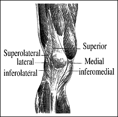
Patients often complain of medial knee pain, with pain occurring with activities such as prolonged sitting with a flexed knee, walking up stairs, or running. Often pain will occur along the medial patella facet or patellar tendon. Most often there is more tightness of the lateral retinaculum. Static patellar glide, medial-to-lateral and lateral-to-medial with the knee flexed 30,� can create pain and limited motion, helping determine where to treat. Tilting of the patella will demonstrate limited motion and tightness, especially when compared with the opposite knee. Palpation of the retinacular tissue will reveal localized tenderness and abnormal tissue sensation. Deep pressure, friction massage, fascial release into the tissue barrier, and sometimes the use of a T-bar are necessary to release the retinacular tissue (Fig. 1). The authors stated3 that most of the patients who benefited from their surgical excising of the painful retinaculum had failed results from previous surgical trauma, blunt injury, or chronic instability.
Reprinted with permission: Kasim N, Fulderson JP. Resection of clinically localized segments of painful retinaculum in the treatment of selected patients with anterior knee pain. Amer J Sports Med 28 (6) 2000.
References
- Voight M, Weider D. Comparative reflex response times of the vastus medialis and the vastus lateralis in normal subjects with extensor mechanism dysfunction. American Journal of Sports Med 19(2):131-137),1991.
- Ahmed AM, Burke DL, Hyder A. Force analysis of the patellar mechanism. J of Orth Res 5:69-85, 1987
- Kasim N, Fulkerson JP. Resection of clinically localized segments of painful retinaculum in the treatment of selected patients with anterior knee pain. Amer J of Sports Med 28(6): 811-814,2000.
- Crossly K, Cowan SM, Fennel KL, McConnell J. Patellar taping: is clinical success supported by scientific evidence? Manual Therapy 5(3): 142-150, 2000.
- Ficat RP, Philippe J, Hungerford DS. Chondromalacia patellae: A system of classification. Clin Orthop 144:43-44,1979.
Warren Hammer,MS,DC,DABCO
Norwalk, Connecticut
Click here for previous articles by Warren Hammer, MS, DC, DABCO.


