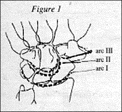
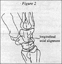
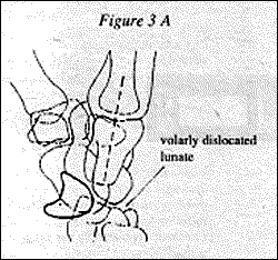
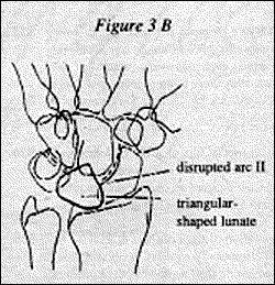
A lunate dislocation on the lateral view is present when the axis of the lunate is angled away from the distal radial surface, while the capitate remains in its normal alignment with the radius. On the PA view, disruption of the arc II is present with lunate dislocations.
Perilunate dislocations can be recognized on the lateral view of the wrist by the dorsal or volar angulation of the longitudinal axis of the capitate, away from its normal central alignment with the lunate and distal radial surface. The lunate in this case remains in articulation with the radius, although there may be some degree of tilt of the lunate.
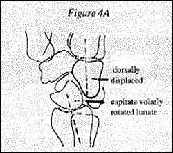
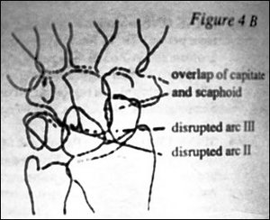
Transscaphoid perilunate dislocations are associated with a fracture of the scaphoid. This type of injury will be demonstrated on the PA view of the wrist; the arc II will be disrupted.
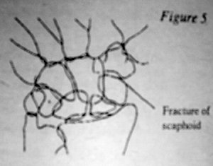
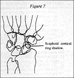
Lastly, the scapholunate dislocation results in injury to the intercarpal ligament that leads to rotary subluxation of the scaphoid. There are two signs that will indicate this type of injury: 1) the Terry-Thomas sign (or maybe David-Letterman for the present) which is widening of the space between the scaphoid and the lunate, which normally measures only 1 mm. 2) the signet-ring sign, which is a cortical ring shadow that is normally not seen on the scaphoid, which is due to rotation of the scaphoid.
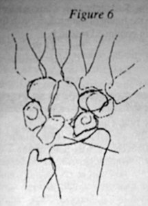
It is extremely important that strict positioning of the wrist be followed, otherwise false positives could become a problem. When obtaining the PA view of the wrist make certain that the wrist is flat against the cassette; making a fist with the hand is a common way to prevent elevation of the wrist. The lateral view should be taken with the wrist in a neutral position in order to evaluate the alignment of the lunate with the rest of the wrist.
References and Diagrams:
Adam Greenspan, Orthopedic Radiology, Gower Medical Publishing 1988.
Click here for more information about Deborah Pate, DC, DACBR.


