


For most patients an AP, lateral and APOM would be the minimal radiographic evaluation I would perform if there were a history of recent trauma and headaches. I don't believe one can get any reasonable idea about what is happening biomechanically at the atlantoaxial region without the APOM. Rotatory fixation is often difficult to evaluate even with an APOM. The view needs to be perfectly aligned to demonstrate the relationship between the dens and the lateral masses.
Patient cooperation is crucial. Once I have the patient's head aligned, I place a sponge to the back of the patient's head. If the sponge falls, I know the patient has moved sometime between my walking away to take the film and my return. Getting the patient to keep the mouth open is also difficult, but sometimes it helps to ask the patient to attempt a yawn. I also increase my kVp by one or two time stations from the AP view, depending on how wide the patient opens his mouth.
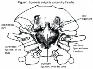
In the APOM, the odontoid should sit equidistant between the articular masses of the atlas. The lateral atlantoaxial (AA) joints are symmetrical in position and configuration. Normally, the principal motion of the AA articulation is rotation, although flexion, extension, lateral translation and vertical approximation also occur to some degree at this level.
It is estimated that approximately half of all rotatory cervical motion occurs at the AA joints; between the anterior arch of the atlas and the odontoid process of the dens; the posterior median AA joint; between the dens and the transverse ligament, encircling the odontoid process and preventing the anterior shift of the atlas on the axis and supporting structures; the paired alar ligaments (AL); and the apical ligament of the dens.
In normal individuals, rotation of the head to either side occurs with the axis of rotation at the anterior aspect of the odontoid process. Rotatory fixation represents the abnormal fixation of atlas and axis in a position of rotation, and has been described in numerous articles, of which those by Fielding and his colleagues should be emphasized (mainly because it was Fielding who developed the four basic classifications of rotatory fixation). They are:
- Type I rotatory fixation without anterior displacement of the atlas.
- Type II rotatory fixation with anterior displacement of the atlas of 3-5 mm, indicating a deficient transverse ligament.
- Type III rotatory fixation with anterior displacement of more than five mm, indicating a ruptured transverse ligament.
- Type IV rotatory fixation with posterior displacement indicating fracture of the dens.
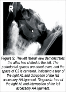
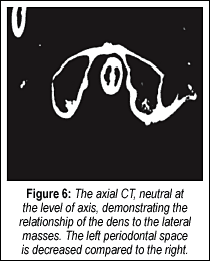
Most of us will only see Type I in our offices. If there is asymmetry between the dens and the lateral masses on the APOM, lateral views in the APOM projection should be performed, especially if there is a history of trauma. Excessive lateral side slippage of C1 on C2 is indicative of rupture or laxity of the accessory ligaments (check ligaments). Cervicocranial axial rotation is primarily limited by the AL, supported by the tectorial membrane, the accessory AA ligaments and the joint capsules.
In axial rotation of C2, the AL on the contralateral side of rotation limits the degree of rotation. When the head is fixed and lateral bending is performed, the atlas will remain fixed with the head, and when the AL are intact, the tension on the contralateral AL ligament will cause the spinous process of C2 to deviate to the contralateral side. The accessory AA ligaments prevent excessive lateral slippage of the lateral masses. As an exercise in biomechanics, let's look at Jackson's description (Figure 2) of ligamentous injuries to the C1-C2 articulations:
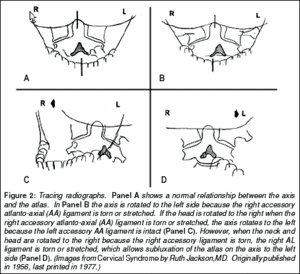
The lateral bending accentuates the stress on these ligaments and allows us to determine if the complex of ligaments is patent before manipulation. I am not making any recommendations on manipulation, but I would recommend before any adjustment is performed that one determine if further damage to the ligament complex is possible.
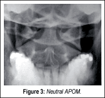
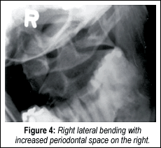
The following case is from Joseph Howe,DC,DACBR, who so kindly wrote the "Upper Cervical Check Ligament Damage" chapter in a book that Sharon Jaeger,DACBR, and I wrote several years ago. See if you can determine the diagnosis. (You may also refer to the captions.)
Deborah Pate,DC,DACBR
San Diego, California
![]()
Click here for more information about Deborah Pate, DC, DACBR.