


Tensegrity - a word combining tension and integrity - is a concept invented by Buckminster Fuller, American architect and inventor known for his geodesic dome design. The basic concept is that biological structures are composed of rigid and tensile structures. Stephen Levin,MD, wrote a splendid article 22 years ago, "Continuous Tension, Discontinuous Compression: A Model for Biomechanical Support of the Body." He contended that "only in failure does the spinal column function as a "stack of blocks," and described how this model applied to the macrostructure.
Dr. Donald Ingber talks about tensegrity and how it applies to basic biological building blocks. His article in Scientific American details the scientific underpinnings of this concept. Here's a quote from Dr. Ingber, on the macrostructure:
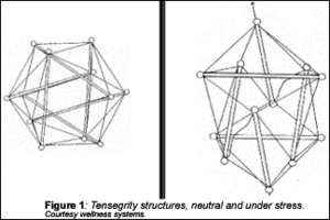
"...an increase in tension in one of the members (within the icosohedral structure) results in increased tension in members throughout the structure - even ones on the opposite side. The principles of tensegrity apply at essentially every detectable size scale in the human body. At the macroscopic level, the 206 bones that constitute our skeleton are pulled up against the force of gravity and stabilized in a vertical form by the pull of tensile muscles, tendons and ligaments. In other words, in the complex tensegrity structure inside every one of us, bones are the compression struts, and muscles, tendons and ligaments (and all interconnected internal fascial structures) are the tensionbearing members."
The relatively rigid structures (the bones) respond to an increased load by compressing, or shortening. This is how bones react when they are traumatized or exposed to prolonged excessive postural stresses. This is the theoretical model underlying the intraosseous restriction in Paul Chauffour's mechanical link concept, and in George Roth's work.
I've corrected intraosseous restrictions for many years, with profound clinical changes. My training in this work started with Dr. Chauffour. My understanding of the underlying principles began to deepen once I took Dr. Roth's tensegrity/matrix repatterning courses. Dr. Gehin put even more of the pieces together for me in understanding why the bones become excessively rigid and compressed.
The connective tissues are always prestressed and under continuous tension. They respond to increased stress with increased tension. All of us who have worked on soft tissue have experienced this. We can release these tensions with our various forms of soft tissue work and adjustments.
The properties of a tensegrity structure are truly remarkable, and make sense as a basis for a new view of biomechanics. I suspect that 20 years from now we will have fully adopted the tensegrity model at the molecular and biomechanical levels.
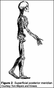
How can we map and assess the entire body using this tensegrity model? Dr. Gehin incorporates a model invented by Rolfer and anatomy teacher Tom Myers,LMT, called "anatomy trains," consisting of a series of "myofascial meridians." Anatomy Trains is a wonderful book for anyone interested in a deeper understanding of the fascial net. Myers outlines the trains (muscles) and stations (the junction points) along these meridians. As a Rolfer, Myers seems to focus his therapies on the MYO, the muscular component of the fascia. Gehin focuses on the intraosseous restrictions along the course of the fascial meridians, and the key junction points.
Let's outline one of these, the superficial posterior meridian. As pictured, it goes along the plantar fascia, up the back of the leg, connecting to ischial tuberosity through the sacroiliac joints, then up the back on either side to the occiput.
When this meridian is shortened, we can see shortened hamstrings; a tight plantar fascia; restrictions within the sacrotuberous ligament; and restrictions affecting the sacroiliac and on up the spine. We all know how to address spinal restrictions, but how often do we look at the sacrotuberous unless we are familiar with the Logan Basic? How many of us consider the plantar fascia only if the patient complains of foot pain? We have to learn to take a broader look and evaluate more completely.
Myers outlines two anterior midline meridians: superficial and deep. These fascial lines, when shortened, pull on the spine. We often need to release anterior restrictions to free up the posterior spinal structures. When we assess spinal conditions, we need to remember that the spine is the backbone, and part of the column of the trunk. A couple of obvious significant structures along the front include the sternum and the sternocostal region, which are often traumatized in motor vehicle accidents by the asymmetric shoulder harness. Deeper anterior structures would include the pericardium (fascial lining of the heart), kidneys, psoas and diaphragm.
Deeper posterior structures are not addressed fully by Myers, but Gehin describes a deep posterior midline fascial meridian that makes perfect sense to any doctor with cranial training. It's the dura going up the spine with its membranous extensions into the skull - the falx cerebri and the tentorium cerebelli. When we release dural tensions in the skull or down the spine, we remove an enormous abnormal tension load from the spine.
We've talked in this series about many of these structures and how to address them. Many others have outlined work on the dura; viscera; psoas; and plantar fascia. What I like about the combination of the tensegrity, anatomy train and myofascial meridian models is the way they remind us to look at the whole system, interconnections and interfaces.
References
- Ingber DE. The architecture of life. Scientific American January 1998. vv.arts.ucla.edu/projects/ingber/ingber.html
- Hammer W. Tensegrity therapy. Dynamic Chiropractic. www.chiroweb.com/archives/19/06/04.html
- Stephen Levin's website on biotensegrity. www.biotensegrity.com/index.html
- Framework classes and articles at www.marchellerdc.com
- George Roth's website, and his articles on application of tensegrity principles. www.wellnesssystems.com
- Tom Myers. Kinesis, anatomy trains website. www.anatomytrains.net
- Myers T. Anatomy trains. Myofascial Meridians for Manual and Movement Therapists, Churchill Livingston, 2001.
- Mechanical link and visceral manipulation seminars (Upledger Institute). www.Upledger.com
Click here for more information about Marc Heller, DC.