


EMG studies have always been considered reliable, but the use of magnetic resonance imaging (MRI) analysis of muscle activity may prove to be more reliable. Flicker et al.,2 studying electromyography for the lumbar muscles, stated that the use of EMGs was complicated due to the particular anatomy of the muscle tested; muscle palpation errors; intersubject/intrasubject variation; and problems in precisely localizing the source of the signal. Magnetic resonance imaging of skeletal muscles may be equal to or better than EMG studies for detecting muscle activity. MRI is based on the concept that exercise produces changes in the amount and distribution of water in skeletal muscle. Magnetic resonance is highly sensitive to changes in water distribution.3 "The water content changes occur mainly due to the production of lactate and other substrates associated with a shift of water out of the vascular into the interstitial and intracellular spaces."4
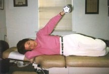
Figure 1: The side-lying abduction exercise.
Horrigan et al.4 used MR when comparing three types of exercises for the rotator cuff muscles: scaption with internal rotation (SIR) "empty the can" or supraspinatus exercise; a side-lying abduction (SLA) exercise in which the patient abducts the arm from the adducted position to 45 degrees abduction and back to the adducted position with the arm in a neutral position (hand facing the body; see Figure 1); and the military press (MP) exercise, in which a patient lifts a barbell overhead with abduction in the scapular plane with the arms in lateral rotation.
The results of the MR study4 showed that of the three exercises studied, the SLA exercise produced the greatest MR signal intensity in the supraspinatus, infraspinatus, subscapularis and deltoid. The SIR at-peak exercise produced the second-highest signal intensity in the trapezius, supraspinatus, subscapularis and infraspinatus and the third-largest intensity in the deltoid. The MP exercise produced the highest signal intensity in the trapezius, second-highest in the deltoid, and third-highest in the supraspinatus and infraspinatus. None of these exercises produced an increase in signal intensity in the teres minor.
One problem when giving the scaption internal rotation exercise for the supraspinatus is that in the 90-degree abduction range, there is the possibility of subacromial impingement, and the patient must be told to refrain from abducting to the ranges that are painful. Horrigan5 feels that abducting the arm to 45 degrees in the side-lying position is effective because the moment arm is the greatest in this position. Therefore, resistance is immediate upon initiation of abduction. Since the glenohumeral/scapulothoracic ratio from 0-25 degrees was 4.1:1, a greater relative emphasis could be placed on the supraspinatus and deltoid. In the study, the SLA exercise was done with dumbbells weighted to 8% of body weight in males and 5% body weight in females.
References
- Townsend H, Jobe FW, Pink M, Perry J. Electromyographic analysis of the glenohumeral muscles during a baseball rehabilitation program. Am J Sports Med 1991;19(3):264-275.
- Flicker PL, Fleckenstein JL, Ferry K, et al. Lumbar muscle usage in chronic low back pain: magnetic resonance image evaluation. Spine 1993; 18(5):582-86.
- Fleckenstein JL, Canby RC, Parkey RW, Peshock RM. Acute effects of exercise on MR imaging of skeletal muscle in normal volunteers. Am J Roentgenol Aug 1988;151(2):231-7.
- Horrigan JM, Shellock FG, Mink JH, Deutsch AL. Magnetic resonance imaging evaluation of muscle usage associated with three exercises for rotator cuff rehabilitation. Med & Sci in Sports & Exer 1999;31(10):1361-66.
- Horrigan. Personal communication, Nov. 1, 1999.
Click here for previous articles by Warren Hammer, MS, DC, DABCO.