


A holistic and educated view of temporomandibular disorders (TMD) reveals how highly functional and neuromuscularly dominant this region is. Okeson verifies this in his guidelines for orofacial pain: "Although TMD was viewed as one syndrome, current research supports the view that TMD is a cluster of related disorders in the masticatory system that have many common symptoms." He goes on to say: "The clinical phenomenon of sensory, motor and autonomic effects resulting from deep pain input are important because referral of pain from the cervical region is common and easily mistaken for masticatory pain."1
"The SCM, although possibly only a responsive muscle, is a common indicator for cervicocranial dysfunction and should signal orofacial investigation,"2 Lewit relates. This article, however, will concentrate on the muscle function and pathology of the orofacial region.
The primary functions of the muscles of mastication include mastication, swallowing and speech. They have secondary roles in respiration and emotional expression. The primary actions of the muscles of mastication involved in mandibular function include elevation (jaw closing); depression (jaw opening); and protrusion (jaw jutting forward). This equates to mandibular opening, closing and lateral shifting, respectively. Lateral excursion and retraction have synergistic action associated with the primary movements listed above. For example, mandibular opening involves inferior depression combined with retraction.
When considering the muscles of mastication and their treatment in relation to TMD, the frequent reply is often unfortunately singular. By far, the near-unanimous attention goes to the lateral pterygoid. The lateral pterygoid certainly warrants attention. It has a role in many aspects of mandibular movement, particularly if it is faulty. To limit application to the lateral pterygoid, however, is not sufficient in treating most TMD.
The lateral pterygoid maintains two divisions, inferior and superior. The superior lateral pterygoid originates from the infratemporal surface of the greater wing of the sphenoid and attaches to the disc (40%) and condyle (60%). Of clinical interest, several recent studies have shown no attachment to the disc in 30% of a population.3 Its functions include stabilization during mandibular elevation and chewing.
The inferior lateral pterygoid originates at the outer surface of the lateral pterygoid plate and attaches to the neck of the condyle. Its primary role is protrusion. With proper activation of the depressors of the mandible, the inferior lateral pterygoid assists in stabilization of rotary movement of the mandible.
The muscles involved with elevation of the mandible are the masseter, temporalis and medial pterygoid. They are postural muscles and are commonly overactive. This corresponds with Janda's discussion of fundamental reflexes and the dominance of jaw closure with elements of fatigue, stress and/or dysfunction.4
Antagonists to the muscles of elevation are the muscles of mandibular depression. They are often inhibited and also house "trigger" and/or "tender" points. The digastric is sighted as the agonist of mandibular depression, with the mylohyoid, platysma and inferior lateral pterygoid acting as synergists.
Discussion of the muscles of mastication must include comment on the hyoid and its reciprocal stabilization with the mandible. Swallow several times and notice that on each occasion your teeth come together. Elevation of the hyoid during swallowing is dependent on mandibular stabilization. In return, efficient mandibular function is greatly dependent on hyoid stabilization and function of its adjacent musculature.
Figure I: Window of the condyle/disc complex of the TMJ. Anteriorly, the superior lateral pterygoid attaches to the articular disc and then joins the inferior lateral pterygoid in attaching to the neck of the condyle. Posteriorly, the articular disc is held by the retrodiscal tissue.
The opening phase of the mandible involves a combination of rotation and translation at the condyle/disc complex (see Figure I). The condyle should rotate on the disc in the inferior joint space for approximately two-thirds of initial opening. The remaining one-third of mandibular opening should occur between the articular disc and the temporal fossa in the superior joint space of the condyle/disc complex. This latter phase would be defined as translation. Many authors agree that rotary movement of the mandible is the most important phase of mandibular function.5 This is most important because it is this phase that establishes proximal stability for the movement of the mandible. The appropriate movement pattern for the mandibular opening would therefore exclude protrusion.
.jpg)
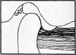
Figure II: Assessing for early protrusion is an excellent screen for muscle imbalance when encountering orofacial pain.
Skill Technique
Mandibular function movement pattern:
- Patient seated or standing
- Patient opens and closes mandible
Qualification:
- Pass/fail
- Fail if mandible protrudes an initial phase of opening
Purpose:
- To identify incoordination of craniomandibular function;
- To determine weakness/inhibition of the depressors of the mandible;
- To determine if lateral pterygoid, masseters, temporalis or medial pterygoid are overactive;
- To screen for functional pathology related to activity limitations with chewing, swallowing, speaking, respiration and emotional expression.
When there is overactivation of the muscles of elevation and inhibition of the muscles of depression, the lateral pterygoid dominates and produces early protrusive activity. Assessing for early protrusion is an excellent screen for muscle imbalance when encountering orofacial pain (see Figure II).6 This early protrusive action places the condyle-disc complex in a vulnerable position, causing abnormal loading locally and to the adjacent head and neck joint structure. Forward head posture and its contributing factors may play a secondary or primary role in the development or perpetuation of this type of functional pathology.
Functional pathology of the orofacial system would typically involve overactivity of the muscles of mandibular elevation and protrusion, inhibition of the muscles of depression, and abnormal loading of the TMJ and cervical spine.
Proper treatment would include relaxation and/or stretching to the overactive muscles and tissues, manipulation and/or mobilization to blocked joints, and facilitation and eventual strengthening to weak and inhibited muscles. Most important and least recognized is the impairment of depression of the mandible and the contributing factors of forward head posture and its related functional pathology.
It is this author's opinion that early aggressive manual therapy could significantly decrease the illnesses and disabilities that produce the staggering statistics associated with TMD and orofacial pain. The practitioner who embraces active rehabilitation and couples it with knowledge and skill in manipulation of the locomotor system will best treat this area. To move the entry point of the patient with TMD to chiropractic and/or physical medicine will require research, documentation and education of the dental and related professions.
References
- Okeson J. Orofacial Pain: Guidelines for Assessment, Diagnosis and Management. Quintessence Books, 1996.
- Lewit K. Manipulative Therapy in Rehabilitation of the Locomotor System, 2nd edition. Butterworth-Heinemann, 1991.
- Naidoo LCD. Lateral pterygoid muscle and its relationship to the meniscus of the temporomandibular joint. Oral Surg Oral Med Oral Pathol Oral Radio Endod 1996;82:4-9.
- Janda V. Some extracranial causes of facial pain. Journ of Prosthetic Dentistry 1986.
- Rocobado M. Arthrokinematics of the temporomandibular joint. In: Clinical Management of Head, Neck and TMJ Pain and Dysfunction. W.B. Saunders Co, 1985.
- Skaggs C. TMJ dysfunction. Journal of Bodywork and Movement Therapy 1997;1(4):198-214.
Clayton D. Skaggs, DC, CCRD
Saint Louis, Missouri