


The unfortunate legacy of line drawing analysis is that it is an illusion. Consider if you will the following scenario. A patient presents to your office with two sets of full spine x-rays taken approximately 90 seconds apart by the same technician and without the patient moving his body.
The case in point is perhaps best illustrated by the following comparison of two lumbar vertebra. These two vertebra are representative of what can be found on a large majority of A-P lumbar films. To the line drawing group the two drawings of the right laterally flexed vertebra will look the same. In reality, they exemplify two distinctly different subluxations and must be diagnosed and treated as such.
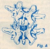
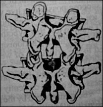
The above illustration has been taken from Kapandji and is for illustration purposes only.
In Figure A (left) the left facet will not close (extend). Thus when the patient is placed in the position of sagittal extension, the right transverse process will become more palpable as a direct result of the shift in the instantaneous axis of motion. The reason is that, as the motion is imparted from superior to inferior through the inferior facets (of the vertebra above), locking on the lamina of the vertebra below (see Bogduk for a more detailed explanation), the right facet (already in the extended position) is carried along into a position of relative hyperextension as a component of the entire spinal coupled-motion phenomenon.
In forward bending, both the transverse processes become more equal in the transverse plane. This occurs as a natural component of the coupled-motions that take place during this motion of transverse rotation and translatory glide. The right facet will open from its relatively extended position to become in line (in a transverse plane) with the left facet in its fixed (flexed) position. This same rational concept can now be used to make a treatment diagnosis. The examining doctor or student, knowing the above information, can also make a positional diagnosis of this dysfunctional vertebral motion unit in a orthogonal coordinate system, that is to say, in three dimensions. The motion unit is fixed in the actions of left extension, left lateral flexion, and left axial rotation. The doctor or student can now select the appropriate adjustive procedure to correct this dysfunctional motion unit taking into consideration that the innate motion of the body is in more than one plane. It should be obvious that this is not a "listing" that can be derived from plane radiography but must come from a clear and accurate understanding of the bodies coupled-motions and a lot of motion palpation practice.
In Figure B the right facet will not open (flex). When the patient is asked to flex in a sagittal plane the right transverse process will more than likely be palpable. This happens because as the motion is from above to inferior, the right facet (which will not flex) is carried along by the coupled spinal motion secondary to the shift in the instantaneous axis of movement to the right side. There is no restriction or fixation of the left facet, therefore the coupled-motion causes a moment about the newly created instantaneous axis and the right transverse becomes prominent to palpation. When the patient attempts the action of retroflexion (spinal extension) the transverse processes become relatively equal in the transverse plane. This is obvious as the left facet is able to move, bringing its transverse process into line with the right or hypomobile one. The motion restriction here is flexion, left lateral flexion and left axial rotation. Clearly this is opposite to the previous picture. Which picture are you going to adjust from: the static model or the one with a sound diagnostic rationale behind it.
The student or doctor will now be able to appreciate the illusion that the two fixations would appear on x-ray to be identical, but on motion analysis they are NOT the same and must be adjusted differently.
Chiropractic, in the years coming, will be coping with managed care. I wonder if we will be ready for it?
Keith Innes, DC
Scarborough, Ontario