


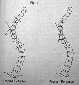
When evaluating patients with a structural scoliosis there are two widely accepted methods of measuring the curve: the Lippman-Cobb and Risser-Ferguson techniques are the most commonly used techniques, but they are not comparable.
A new technique for measuring the degree of scoliosis was introduced in 1978 by Adam Greenspan and is more accurate in measuring the deviation of each involved vertebra. This technique, called the "scoliotic index," measures the deviation of each involved vertebra from the vertical spinal line, as determined by a point at the center of the vertebra immediately above the upper end-vertebra of the curve, and the center of the vertebra immediately below the lower end-vertebra. This technique is particularly useful when evaluating short segments or minimal curvatures, which are often difficult to measure with the currently accepted methods. (Fig. 3)
In addition to measuring the scoliotic curvature, the radiographic evaluation of scoliosis also requires the determination of other factors, including measurement of the degree of rotation of the vertebrae and the skeletal maturity of the patient. The measurement of the degree of rotation of the vertebrae of the involved segments can be obtained by either of two methods. Cobb's technique for grading rotation uses the position of the spinous process as a point of reference. Normally the spinous process should appear in the center of the vertebral body if there is no rotation. As the degree of rotation increases, the spinous processes migrate toward the convexity of the curve. (Fig. 4) Moe's method for grading rotation uses the symmetry of the pedicles as point of reference, with the migration of the pedicles toward the convexity of the curve determining the degree of vertebral rotation. (Fig. 5)
The final factor in the evaluation of scoliosis is the determination of skeletal maturity. This is important for both the prognosis and treatment of scoliosis, particularly the idiopathic type, since there is a potential for significant progression of the degree of curvature, as long as skeletal maturity has not been reached. Skeletal age can be determined by comparison of a radiograph of a patient's hand, with the standards for different ages available in radiographic atlases. It can also be assessed by radiographic observation of the status of ossification of the apophysis of the vertebral ring, or from the status of the ossification of the iliac apophysis. (Fig. 6)1
It is generally accepted that any curve under 50 degrees should be treated conservatively. Treatment of curve should consist of chiropractic care and adjunctive exercises. If the curve, however, is over 50 degrees, orthopedic consultation should be sought for the benefit of the patient and for malpractice protection.
Reference:
1. The information for this article was obtained from Orthopedic Radiology by Adam Greenspan, J.B. Lippincott, 1988.
Click here for more information about Deborah Pate, DC, DACBR.