


Sacro-occipital technique (SOT) teaches that the anterior and posterior aspects of the SI joint are completely different in both their anatomy and function. The posterior aspect has hyaline cartilage and is a weight-bearing joint that should not have much, if any, motion. On the other hand, the anterior aspect has a synovial bed, which allows for the joint nutation and whose motion purportedly aids in cerebrospinal fluid (CSF) mixing in a cranialward direction.
One aspect of the SI joint, when mobile, would be dysfunctional (e.g., SOT's category two), and one aspect of the SI joint when too "stable," would be dysfunctional (e.g., SOT's category one). It seems as if the chiropractic profession is only looking at the SI joint as being fixated, which seems to me to be fixated thinking. The first step in going beyond this fixated thinking would be differentiating between a hypermobile SI (category two) and a fixated one (category one). Is the dysfunction associated with the posterior weight-bearing aspect or is the anterior portion associated with normal nutation? See Fig. 1 below.
The second step is differentiating between a fixated SI joint secondary to an anterior joint dysrelationship, such as a pelvic torsion,1 or a fixated SI joint secondary to a posterior hypermobile joint causing, neuromuscularly speaking, a "splinting" due to increased nociception and local muscle hyperfaciliation, leading to increased myofascial tension.2 This increased tension will simulate an actual osseously fixated joint, but is fixated muscularly as a guarding and protective mechanism.
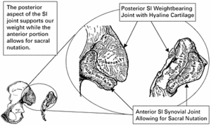 Figure 1: This illustrates the difference in anatomical structure of the anterior and posterior aspects of the sacroiliac joint.
SOT is an indicator-based system that uses the patient's report of pain and tension at specific locations to guide treatment, since if the "indicators" worsen, the treatment would need to be modified. If they improve, the doctor knows they are going down the right path. The indicators help guide the doctor to realize the patient's progress independent of what a patient might say about his or her condition. When the indicators and symptoms are not congruent, that indicates the need for more extensive diagnostic protocols.3
Figure 1: This illustrates the difference in anatomical structure of the anterior and posterior aspects of the sacroiliac joint.
SOT is an indicator-based system that uses the patient's report of pain and tension at specific locations to guide treatment, since if the "indicators" worsen, the treatment would need to be modified. If they improve, the doctor knows they are going down the right path. The indicators help guide the doctor to realize the patient's progress independent of what a patient might say about his or her condition. When the indicators and symptoms are not congruent, that indicates the need for more extensive diagnostic protocols.3
A concept that DeJarnette, the developer of SOT, determined was that there can be aspects of both an anterior and posterior dysfunction in a SI joint. However, if there is even a small aspect of posterior hypermobility, that usually should be the focus of treatment prior to addressing any anterior SI joint fixation with the purpose of ultimately increasing joint motion. In essence, DeJarnette determined that body stability associated with weight-bearing stresses generally supersede the need to maintain normal sacral nutation.
The treatment of the SI joints involves eliminating myofascial influences that might be affecting pelvic torsion or rotation, as well as any possible confounder associated with a leg-length discrepancy. Each of the SOT categories has a particular manner of determining leg-length inequality, which will guide treatment. Generally with SOT, the treatment uses pelvic blocks, but it is mostly due to preference, effectiveness and its low force. As long as the doctor can balance the indicators associated with anterior or posterior SI joint dysfunction, DeJarnette didn't really care what method of treatment was rendered.
Most category indictors are related to increased muscle tension, places of increased pain in specific regions associated with each individual category and related altered body function (Fig. 2). One way of evaluating whether appropriate treatment is being rendered is the lessening of pain or tension at those specific points, as well as balanced function of the indictors. Both posterior SI joint hypermobility (category two) and anterior SI joint fixation (category one) will have their own set of places of pain and tension, which resolve with appropriate care.
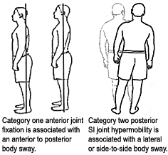 Figure 2: Body sway in the standing posture – differentiating anterior versus posterior sacroiliac joint dysfunction.
Specific Palpatory Pain Indicators
Figure 2: Body sway in the standing posture – differentiating anterior versus posterior sacroiliac joint dysfunction.
Specific Palpatory Pain Indicators
Generally, there are some ways to differentiate between joint fixation versus hypermobility. With joint hypermobility, you will tend to find increased sensitivity anywhere along the inguinal ligament (located bilaterally between the ASIS and the pubic bone), along with either medial (sartorius insertion) or lateral knee (tensor fascia lata insertion) pain on either or both legs (Fig. 3). With a category one, there commonly are places of pain at the region where the piriformis and gluteus medius cross, as well as at the lumbodorsal fascia just lateral to the L4/5 region (Fig. 3). Category one pelvic torsion tends to cause whole-body axial torsion, while category two SI joint dysfunction will have aspects of whole-body torsion as well as lateralized dysfunctions. Category two SI joint instability tends to be unilateral, and the body's kinematic chain, in its inability to translate gravity, will accommodate by having multiple lateral postural unlevelings, from the pelvis to the head.
Category two presentations can have radiating pain along the anterior lateral thigh and multiple related joint dysfunctions at the knees, shoulders and TMJ. Category one tends to cause symmetrical joint dysfunction and is more commonly associated with generalized neurological dysfunction, lowered pain and body function thresholds, and somatovisceral/viscerosomatic (mimicry) involvement.
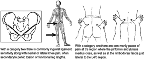 Figure 3: Palpatory referred muscle and pain points – differentiating between anterior and posterior SI joint dysfunction.
1st Rib - Scalenus Muscle Tension
Figure 3: Palpatory referred muscle and pain points – differentiating between anterior and posterior SI joint dysfunction.
1st Rib - Scalenus Muscle Tension
The 1st rib is different than each of the other 11 ribs in that its joint is based in hyaline cartilage, while the other ribs are synovial-type joints. While the other 11 ribs move with respiration, the 1st rib is supportive and does not. Since the body is a holographic kinematic chain, dysfunction in the SI joint will be represented throughout the body. One place offering information is the 1st rib costovertebral junction. Increased motion in the 1st rib will tend to lead to increased scalenus muscle tension as the muscle attempts to support and splint. DeJarnette has found that a category one tends to affect the 1st rib/scalenus bilaterally and a category two affects the 1st rib/scalenus unilaterally (Fig. 4).
SOT postulates that the scalene/first rib4 area relates to category two due to visual and vestibular righting reflexes,5,6 an attempt of the body to maintain an upright posture when challenged by the assymetrical joint-loading of one SI joint in the category two patient.
Treating SI Joint Hypermobility (Category Two)
Category two SI joint hypermobility is treated predominately in a supine position2,7 with pelvic blocks placed according to pelvic torsion presentation. Pelvic torsion can be determined by multiple methods, but evaluation is not performed until all related myofascial influences affecting the pelvis have been released. Most commonly, the main compensatory factor affecting pelvic torsion is imbalance of the iliopsoas/quadratus lumborum muscles, piriformis/gluteus medius muscles, and upper cervical vertebra. SOT uses various methods to diagnose this restriction relating to palpation of the tissues, evaluating muscle function, such as the "over-the-head arm check," which evaluates the ability of the rib cage to lift from the pelvis. SOT treatment first addresses the myofacial issues. Once there is balanced flexibility, strength, and function of the specific muscles, proceed to the direct treatment of the SI joint.
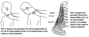 Figure 4: Palpatory pain and function at thoracic inlet – differentiating between anterior and posterior SI joint dysfunction.
Category two leg lengths are determined by having the patient abduct his or her legs (15 inches apart) against resistance. After a few seconds of doctor resistance, the patient relaxes while the doctor maintains traction on the legs while determining if one medial malleoli is superior or inferior to the other. If the pelvis is imbalanced, pelvic blocks will be used to reduce pelvic torsion and compress the posterior hypermobile SI joint.
Figure 4: Palpatory pain and function at thoracic inlet – differentiating between anterior and posterior SI joint dysfunction.
Category two leg lengths are determined by having the patient abduct his or her legs (15 inches apart) against resistance. After a few seconds of doctor resistance, the patient relaxes while the doctor maintains traction on the legs while determining if one medial malleoli is superior or inferior to the other. If the pelvis is imbalanced, pelvic blocks will be used to reduce pelvic torsion and compress the posterior hypermobile SI joint.
Traditionally, the main test of a category two is the arm fossa test. This tests a patient's ability to respond by holding his or her arm in a consistent position as the doctor contacts the inguinal ligament. While the test has had some degree of acceptance as a SI joint evaluative test,8 it does take time to learn and perfect in order to get consistent results. For that reason, this presentation will mostly focus on easy-to-monitor-and-test indicators such as palpation for pain and increased tension (Fig. 5). If the patient is determined to have a category two presentation and the blocks are placed properly, inguinal ligament sensitivity or medial/lateral knee pain (if present) will begin to subside within 30-60 seconds of block placement. Within one to two minutes, the anterior scalenus muscle tension tends to become more symmetrical and less sensitive.
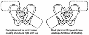 Figure 5: Supine pelvic block treatment. Block placement for category two – posterior SI joint hypermobility.
Sometimes it will take a couple of visits, but the standing sway pattern will begin to balance and anterior/lateral thigh pain will begin to subside. When the patient's indicators are not resolving, there are a few possibilities, such as concomitant SI sprain with sacral segment posterior translation, piriformis muscle syndrome secondary to anterior sacral translation, L4/L5 discopathy secondary to iliolumbar ligament dysfunction, and descending TMJ dysfunctional righting mechanisms.9-11
Figure 5: Supine pelvic block treatment. Block placement for category two – posterior SI joint hypermobility.
Sometimes it will take a couple of visits, but the standing sway pattern will begin to balance and anterior/lateral thigh pain will begin to subside. When the patient's indicators are not resolving, there are a few possibilities, such as concomitant SI sprain with sacral segment posterior translation, piriformis muscle syndrome secondary to anterior sacral translation, L4/L5 discopathy secondary to iliolumbar ligament dysfunction, and descending TMJ dysfunctional righting mechanisms.9-11
There are varying levels of category two syndromes associated with significant joint and articular capsule spraining, and other less serious conditions related to posterior joint-increased proprioception irritation and decreased nociception thresholds. Generally, the less severe condition will respond relatively quickly, but the patient will need to be cautioned to resist the temptation to do too much joint motion or joint loading. Healing the joint usually takes around four to six weeks, but with fewer traumas, the joint can heal quicker; with chronically sprained joints, it can take longer. With a chronically sprained joint, the patient will need to perform rehabilitative exercises such as straight prone leg raises, swimming, walking and other types of activities that can stimulate posterior joint circulation to increase ligamentous strength and healing.
Note: All illustrations included in this article are reproduced with permission from Robert Monk, DC.
References
- Cooperstein R, Lisi A. Pelvic torsion: anatomic considerations, construct validity, and chiropractic examination procedures. pTopics in Clinical Chiropractic, Sept. 2000;7(3):38-49.
- Knutson G, The sacroiliac sprain: neuromuscular reactions, diagnosis and treatment with pelvic blocking. Journal of the American Chiropractic Association, Aug. 2004,41(8):32-9.
- Blum CL. Incongruent sacro-occipital technique examination findings: two unusual case histories. Proceedings of the ACC Conference IX. Journal of Chiropractic Education Spr 2002; 16(1):67.
- Blum C, Curl D. The relationship between sacro-occipital technique and sphenobasilar balance. Part one: the key continuities. Chiropractic Technique 1998;10(3):101-107.
- Bronstein A, Guerraz M. Visual-vestibular control of posture and gait: physiological mechanisms and disorders. Curr Opin Neurol, Feb. 1999;12(1):5-11.
- Morningstar M, Pettibon B, Schlappi H, Schlappi M, Ireland T. Reflex control of the spine and posture: a review of the literature from a chiropractic perspective. Chiro & Osteop 2005;13:13-16.
- Cooperstein R. Padded wedges for lumbopelvic mechanical analysis. Journal of the American Chiropractic Association, Oct. 2000:24-6.
- Hestk L, Leboeuf-Yde C. Are chiropractic tests for the lumbopelvic spine reliable and valid? A systematic critical literature review. Journal of Manipulative and Physiological Therapeutics, May 2000;23:258-75.
- Chinappi, AS, Getzoff, H. Chiropractic/dental co-treatment of lumbosacral pain with temporomandibular joint involvement. J Manipulative Physiol Ther, Nov/Dec 1996;19(9):607-12.
- Chinappi AS Jr; Getzoff H. The dental-chiropractic co-treatment of structural disorders of the jaw and temporomandibular joint dysfunction. J Manipulative Physiol Ther, Sept. 1995;18(7):476-81.
- Blum C. A chiropractic perspective of dental occlusion's affect on posture. Poster presentation: Proceedings of the ACC Conference XI, Journal of Chiropractic Education Spr 2004;18(1):38.
Dr. Charles Blum, a graduate of Cleveland Chiropractic College - Los Angeles, is a past president of SOTO-USA and the organization's representative to the AATMD.