


The laser was theorized by Einstein in 19171 and invented by Maiman in 1960.2 Its unique property of light waves being coherent in space and in time led many to theorize that it could be a damaging form of electromagnetic radiation.
Thus the field of laser therapy was born and now, 42 years later, therapeutic lasers are gaining acceptance in the chiropractic profession for the treatment of chronic pain, sports injuries, musculoskeletal conditions and more. While exhibiting at a chiropractic trade show a couple years ago, a wily old doctor asked me, "How do you know you're not causing cancer?" which is an excellent question. Let's talk about laser and its effects on tissue.
Before we answer the question of whether laser therapy can damage tissue, let's review some basic physics terms applicable to laser therapy. Power is measured in Watts and is the time rate of energy delivery. Energy is measured in Joules, and time in seconds. Energy density, also known as dosage, measures the amount of energy applied per unit area in Joules per square centimeter. Power density, or irradiance, is measured in Watts per square centimeter.
| Table 1. Physics Terms and Units for Therapy Lasers | ||
| Term | Description | Unit of Measurement |
| Power | Time rate of energy delivery | Watts, W |
| Energy | Ability to do work | Joules, J |
| Wavelength | Distance between like points of a wave | nanometers, nm |
| Energy Density | Dosage | Joules per square centimeter, J/cm2 |
| Power Density | Irradiance | Watts per square centimeter, W/cm2 |
Wavelength is a measure of the distance between adjacent points on a light wave and is expressed in nanometers (nm). Therapy lasers use visible and near-infrared light in the range of 635 nm to 1,064 nm. Wavelength determines how deep laser energy penetrates into tissue.3 The energy of individual photons is inversely proportional to the wavelength. Photons of ultraviolet light carry higher energy than photons of infrared light. (See Figure 1)
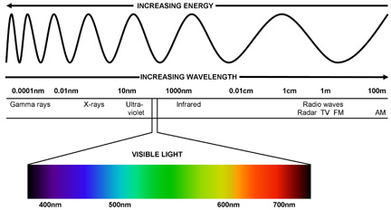
All living matter is composed of molecules which are made of atoms held together by electron bonds. Ionizing radiation is energetic enough to break those bonds. High-energy photons such as gamma rays, X-rays and ultraviolet rays are capable of ionizing molecules, causing damage to cell components and DNA. Lower energy photons cannot ionize molecules, instead inducing vibration, rotation and translation within the molecule. These low-energy transfers are not capable of damaging cells or DNA.
There is a very clear wavelength limit at which the energy of individual photons becomes high enough to ionize molecules: 320 nm, which sunbathers know as UVb.4 All electromagnetic radiation with a wavelength shorter than 320 nm (to the left of UV in Figure 1) is capable of ionizing living matter and causing cancer or other cellular damage.
 Figure 2: Map of laser-tissue interactions showing power density versus exposure time.7
Power density, or irradiance, measures the concentration of the laser's "brightness." Low-power densities produce beneficial photochemical and photobiological effects in tissue. Higher power densities result in thermal interactions, whereby the local temperature is elevated significantly5.
Figure 2: Map of laser-tissue interactions showing power density versus exposure time.7
Power density, or irradiance, measures the concentration of the laser's "brightness." Low-power densities produce beneficial photochemical and photobiological effects in tissue. Higher power densities result in thermal interactions, whereby the local temperature is elevated significantly5.
In laser-tissue interactions the term thermal does not refer to mild warming of a few degrees, but to increases in tissue temperature above 60 degrees C, producing the effects of coagulation, vaporization, carbonization and melting.6
To calculate the power density of a therapy laser, divide the average power delivered by the spot size of the laser. For example, a 500 milliwatt (0.5 W) laser with a 1 centimeter diameter spot size would produce a power density of 0.6 W/cm2, and a 10 Watt laser with a 2.5 centimeter spot size produces a power density of 2 W/cm2. Therapy lasers deliver output powers ranging from 0.005 W to 12 W, and when used as intended operate at power densities of 2 W/cm2 or less.
Figure 2 shows the five basic types of laser-tissue interactions plotted on a chart of power density versus exposure time. Note that the axes are logarithmic, where 106 = 1,000,000. The zone of action for therapy lasers is at the lower right, where power densities are less than 10 W/cm2. Once again, in the description of laser-tissue interactions the term thermal interaction refers to those interactions that produce a significant (>60 degrees C) rise in temperature, not the mild increases of a few degrees that can happen with laser therapy treatments. (See Table 2 below)
| Table 2. Thermal Effects of Laser Radiation8 | ||
| Temperature | Biological Effect | |
| Celsius | Fahrenheit | |
| 37 | 99 | Normal |
| 39 | 102 | (Typical maximum temperature in region of Class IV therapy laser treatment) |
| 40 | 104 | (Maximum suggested temperature for hot tub) |
| 45 | 113 | Hyperthermia |
| 50 | 122 | Reduction in enzyme activity, cell immobility |
| 60 | 140 | Denaturation of proteins and collagen; coagulation |
| 80 | 176 | Permeabilization of membranes |
| 100 | 212 | Vaporization; thermal decomposition (ablation) |
| > 100 | > 212 | Carbonization |
| > 300 | > 572 | Melting |
Energy density, or dosage, measures the total amount of energy delivered per unit area and is expressed in J/cm2. Too low a dose is ineffective, just the right dose produces the desired effect, and too high a dose can be biosuppressive. What is the right dose? Biostimulation has been reported in studies with dose ranges from 0.001 J/cm2 to 10 J/cm2 and higher.9
 Figure 3: Dose-response curve for laser therapy.
Determining the appropriate treatment dose is a complicated issue and depends on many factors such as wavelength, power density, condition, treatment technique and more. In their text Laser Therapy: Clinical Practice and Scientific Background, Drs. Jan Tuner and Lars Hode suggest dosages from 1.0 to 10.0 J/cm2 for the treatment of superficial or deep pain conditions.10
Figure 3: Dose-response curve for laser therapy.
Determining the appropriate treatment dose is a complicated issue and depends on many factors such as wavelength, power density, condition, treatment technique and more. In their text Laser Therapy: Clinical Practice and Scientific Background, Drs. Jan Tuner and Lars Hode suggest dosages from 1.0 to 10.0 J/cm2 for the treatment of superficial or deep pain conditions.10
How much dosage is too much? One study has reported irreversible cellular and genetic damage as a result of high-energy densities.11 In this particular study a dose of 16 J/cm2 was delivered to naked cells, whereas the maximum recommended dose in the treatment of deep pain is 10 J/cm2. It should obvious that there is a great difference between irradiating in vitro cultures just a few cell layers thick, and living human patients.12
So, can laser therapy damage tissue? Yes it can, under two conditions: it is of sufficient intensity to burn (high power density); or it contains high-energy photons (short wavelength).13
As indicated, therapy lasers deliver power densities that produce photochemical and photobiological responses in tissue at levels significantly lower than those needed to damage tissue. The dosages recommended for laser therapy are far below those needed to damage cell cultures. Also, with their wavelength range from 635 nm to 1,064 nm, therapy lasers produce photons that do not ionize living matter. So, laser therapy does not cause cancer, or damage to skin, tissues or DNA, because therapy laser light is non-ionizing and is delivered at dosages far below those needed to cause damage.
References
- "Zur Quantentheorie der Strahlung" ("On the Quantum Theory of Radiation"). Physika Zeitschrift, Volume 18, 1917:121-128.
- Jeff Hecht. Beam: The Race to Make the Laser. Oxford University Press, New York:3.
- Markolf H. Niemz, Laser-Tissue Interactions: Fundamentals and Applications. University of Heidelberg, Springer-Verlag Berlin Heidelberg, 2007:3.
- Tuner J, Hode L. Laser Therapy: Clinical Practice and Scientific Background. Prima Books, 2002: 4.
- Markolf H, Op Cit, 47.
- Markolf H, Op Cit, 58-59.
- Markolf H, Op Cit, 46.
- Based on Markolf, page 78, with other values added for reference.
- Tuner and Hode, Op Cit, 72.
- Tuner and Hode, Op Cit, 73.
- Houreld NN, Abrahamse H. In vitro exposure of wounded diabetic fibroblast cells to a helium-neon laser at 5 and 16 J/cm2. Photomedicine and Laser Surgery, 2007;25(2):78-84.
- Tuner and Hode, Op Cit, 72.
- Tuner and Hode, Op Cit, 4.
Part 1 of this article series appeared in the Jan. 1, 2009 issue of DC; part 2 appeared in the July 1 issue.
Dr. Phil Harrington is a certified medical laser safety officer and serves on the subcommittee reviewing the ANSI Standards for Safe Use of Lasers in Health Care Facilities. He is a 1996 Palmer graduate and also holds a bachelor's degree in physics. He is the medical director, clinical manager and laser safety officer for Summus Medical Laser (www.summuslaser.com).