


Unfortunately, knee pain and dysfunction are common. It is not surprising that of all the areas treated by orthopedic surgeons, the knee is the most commonly injured joint, representing a whopping 26 percent of orthopedic business.1 In my estimation, the average chiropractor will run into at least one knee case on a daily basis.
Knee Anatomy 101
In order to determine how to classify and treat these injuries, a brief review of the relevant anatomy is in order. The knee is a hinge-type synovial joint composed of three functional compartments. The patellofemoral articulation, which consists of the patella or "kneecap," and the patellar groove on the front of the femur through which it slides; and the medial and lateral tibiofemoral condyles, which connect the femur with the tibia. The joint is bathed in synovial fluid, which is contained inside the joint capsule.
 The pair of tibial condyles is separated by the intercondylar eminence, composed of a lateral and a medial tubercle. The patella is inserted into the thin anterior wall of the joint capsule. Its posterior surface includes a lateral and a medial articular surface, both of which communicate with the patella surface, which unites the two femoral condyles on the anterior side of the bone's distal end.
When patients present to a doctor's or therapist's office, they are usually doing so in response to some sort of injury or pain they are experiencing. In our society, we have been raised with the allopathic medical mentality, "If it hurts, it's a problem; if it doesn't hurt, you are fine." It is because of this type of thinking that patients focus heavily on a specific mechanism of injury or single incident that may have caused this injury to occur.
Misalignment Patterns
The knee can certainly be a complex structure, and there are orthopedic and medical specialists who deal with this area exclusively. These medical professionals are the ones whose answers usually include medication, cortisone injections, surgical repair / clean-up or sending the patient to physical therapy.
In certain cases of severe injury or trauma, these may, in fact, be appropriate. There is a time and a place for all types of treatment. But as chiropractors, we typically think outside the box. Understanding biomechanics and how adjacent joints affect each other is what we do on a daily basis. It is our sense of how things move in the body that gives us the ability to really understand what the body is experiencing.
No matter what type of practice you have and no matter which technique(s) you use, you see subluxation or misalignment patterns emerge. These patterns allow you to find out what is happening in the patient. These patterns also act as a roadmap for your treatment and rehabilitation.
When all conditions are healthy and "normal," standing and gait make the ground force enter the center of our heel and move into the forefoot. From here, the energy wave goes up through the center of the tibiotalar joint and the tibiofemoral joint. Forces are distributed equally and through the portions of the joint that are made to cushion and dissipate stress or shock.
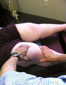 Figure 1: Setup for the medial tibia / femur adjustment.
In my experience, only about 10 percent of the general patient population displays healthy feet that have well-supported arches. Their bodies will take time to display signs of wear and tear, as they usually can take stress in their joints better. How the foot functions is directly related to how well the knee will function. For example, a study measured hyperpronation of the foot and ankle complex with the navicular drop test. Subjects who had ACL injuries had greater navicular drop test scores than non-injured subjects. This suggests hyperpronation of the foot and ankle complex may increase the risk of injury to the ACL.2
Figure 1: Setup for the medial tibia / femur adjustment.
In my experience, only about 10 percent of the general patient population displays healthy feet that have well-supported arches. Their bodies will take time to display signs of wear and tear, as they usually can take stress in their joints better. How the foot functions is directly related to how well the knee will function. For example, a study measured hyperpronation of the foot and ankle complex with the navicular drop test. Subjects who had ACL injuries had greater navicular drop test scores than non-injured subjects. This suggests hyperpronation of the foot and ankle complex may increase the risk of injury to the ACL.2
Overpronation is common, affecting around 80 percent of the population. You might remember what type of stress pattern this sets up for the body. When we pronate and our feet drop down toward the floor, there is a general collapse of the three weight-bearing arches of the foot. The dropping of the arches places pressure on the medial ankle and tibia.
As the foot drops, the tibia moves internally (medially) and this, in turn, causes the femur to move likewise. The worse the pronation or flattening of the foot, the more severe the internal rotational forces are placed on the tibia and femur bones. If you stand up right now and push your feet downward to the floor, you can feel the inward rotation of these bones and the subsequent pressure on the medial knee region. This standing exercise is a powerful way for you and your patients to understand the effect the feet have on the knees and the rest of the body.
Furthermore, the patellae are pulled medially; that is why we often see the "knock-kneed" appearance of a classic overpronator. Even in an average circumstance, the overpronation that exists (even in mild cases) is enough to begin putting stress and microtrauma on the knee and its structures.
With this distortion pattern emanating from the feet and moving up through the ankles to the knees, it makes sense why we see so many common knee-region injuries in our practice. Anteromedial knee pain, shin splints, ACL stress / tears, medial meniscus degeneration, medial collateral ligament damage, partial or total knee replacements, Osgood-Schlatter and patellar tracking disorders are recurring issues you have probably seen and are currently seeing in your patients.
Have you thought about the cause of these conditions? Researchers Rothbart and Estabrook found that a chondromalacia model explained "the pathomechanical events associated with oblique tracking patellar syndrome. The authors suggest that excessive pronation is the causative factor directing asynchronous rotation between the shank and femur."3
When we think about the percentages, since 80 percent of the population overpronates, about eight of 10 patients will display medially rotated tibias, femurs and patellae. Understanding this concept allows us to adjust these bones quite effectively. By performing light- to medium-force adjusting techniques to de-rotate the leg bones, we can restore better bony alignment. Here are a few examples:
Adjusting the Knee
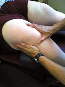 Figure 2: Setup for the medial / inferior patella adjustment.
With the knee in slight flexion on the table, contact the proximal tibia with one hand and the distal femur bone with the other. Then use gentle medial to lateral pre-stress to end range and add a very light rotatory thrust on the tibia to gently decompress the medial knee region. Thus, the medially rotated tibia and femur bones are moved into external rotation.
Figure 2: Setup for the medial / inferior patella adjustment.
With the knee in slight flexion on the table, contact the proximal tibia with one hand and the distal femur bone with the other. Then use gentle medial to lateral pre-stress to end range and add a very light rotatory thrust on the tibia to gently decompress the medial knee region. Thus, the medially rotated tibia and femur bones are moved into external rotation.
The patella, which has moved medially and inferiorly, can be moved as well. With the knee again in slight flexion, take your inside hand and cup the inferior, medial border of the patella with the thumb web. The stabilizing hand is placed on the lateral side of the knee for support. Then a slight pressure is placed in the superior, lateral direction to gently shift the patella into better alignment.
The fibular head can also be considered here, as it tends to move superiorly/laterally in response to the excessive foot pronation. This maneuver can usually be accomplished by grabbing the fibular head with the thumb, index and middle fingers, and applying slight inferior-to-anterior force.
Remember that for any of the adjustments described above, there are variations we do not have the space to discuss. Also, you can utilize drop table or spring-loaded instrument adjusting as well. Just remember your patterns of misalignment and then have fun correcting the bony alignment however you see fit.
I must make mention of the 3-5 percent of the population that falls into the true classification of excessive supinators or oversupinators. These people actually roll to the outside of their feet, and usually have a high medial arch with low to flat lateral and transverse arches. These individuals tend to display lateral knee pain with their tibia and femur bones in excessive external rotation. In essence, correcting these bones is in the opposite direction of the overpronators.
Regardless of the pronation or supination pattern, adjusting the feet and ankles is important as well. Once the adjustments have been made, it's then important to help stabilize the joint. Here are some tools that can be utilized:
Joint Stabilization Strategies Following Your Adjustments
- Elastic sports tape: This is a simple tool that can have a great impact on the area needing support.
- Knee bracing: This adds much-needed support to the knee while allowing for range of motion in most activities.
- Stabilizing foot orthotics: This addresses the arches of the foot, which as discussed earlier, will impact the kinetic chain and help to stabilize and give the knee a more balanced foundation.
- Proper shoe type: This is overlooked by most patients and is the easiest way to properly support their feet / knees. Discuss what shoe types are appropriate for specific activities on a individualized basis.
The knee is an amazing joint of the lower extremity, but it is one in which pain / dysfunction are common. Pain can often be traced back to the feet and ankles. Although we should never overlook the possibility of a traumatic or acute-onset injury, step back, think and see if you can draw some connections as to what the patient is feeling and what biomechanical patterns exist from the feet upward. This may provide substantial insight into not only the problem, but also how to treat it effectively.
References
- How the Knee Works, Knee Pain, Wear-and-Tear Injuries, ACL Injuries. The Lower Extremity Center at The Orthopedic Institute: http://orthopedicinstitutesf.com/faq/knee.html.
- Beckett M, Massie D, Bowers K, Stoll D. Incidence of hyperpronation in the ACL injured knee: a clinical perspective. Journal of Athletic Training, 1992;27(1):58-62.
- Rothbart BA, Estabrook,L. Excessive pronation: a major biomechanical determinant in the development of chondromalacia and pelvic lists. J Manip Physiol Ther, 1988 Oct;11(5):373-9.
This is the third in a series of six articles dedicated to the extremities, covering clinical discussions from the feet to the ankles, knees, hips, hands / wrists and elbows to the shoulders. Part 1 appeared in the Feb. 12, 2012 issue; part 2 ran in the April 22 issue.
Dr. Kevin Wong, earned a BS in exercise physiology from the University of California – Davis and his DC degree from Palmer Chiropractic College West. He practices in Orinda, Calif., and serves the Lamorinda, Berkeley, Walnut Creek and many other East San Francisco Bay Area communities. He is an expert on foot analysis, walking and standing postures, and orthotics, and lectures nationwide on spinal and extremity adjusting.