


Throughout our history, many chiropractors have assumed that segmental dysfunction in the axial skeleton involves osseous malposition at a motor unit. The "bone out of place" concept has been used to explain several clinical phenomena including nerve entrapment; palpatory edema and point tenderness; restricted or altered range of motion including joint fixation; and periarticular soft-tissue involvement with muscle tension, spasm or flaccidity.
Clinicians use diagnostic imaging, motion and static palpatory findings, and a wide range of orthopedic and neurological tests, to corroborate the osseous misalignment subluxation model. Recent studies challenge some of these assumptions, especially with regard to sacroiliac joint (SIJ) involvement.
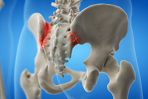 Vleeming, for example, argues that "[A] dysfunctional SIJ is normally not related to a subluxated position of the joint, but to increased or decreased compression/force closure due to asymmetric forces acting on the joint."1 Therefore, what we see on X-ray is likely the result of soft-tissue imbalance (force closure), rather than the osseous distortion seen on X-ray.
Radiographic Evidence in Question?
Radiographic evidence of at least one form of osseous misalignment and malposition at the SIJ has also been called into question. Several anatomical studies show that the SIJs lie obliquely at an angle to the sagittal plane. In the standing position, the cranial portion of the joint is primarily vertical in a parasagittal plane, but with the keystone-like bony anatomy, the sacrum is "wedged cranially and dorsally into the ilia within the pelvic ring."2 The rigidity and relative immobility, under normal stresses, are an example of "form closure" described by Vleeming in subsequent studies.
Consequently, there is little to no significant room for the sacrum to achieve osseous displacement relative to the ilia. While the Gonstead method of plain-film analysis suggests axial rotation (θy) of the ilium relative to the sacrum with an IN or EX listing of the ilium, a study by Weinert indicates such an observation is most likely an artifact of positioning.
Even small degrees of axial rotation of the patient's pelvis may create an optical illusion of SIJ misalignment. Therefore, Weinert concludes, "Significant associations between [patient] axial rotation and chiropractic pelvic line analyses obtained from radiographs were found ... raising questions of the clinical usefulness of pelvis radiographic line analysis of iliac and sacral rotation."10 Positioning artifact may create "IN ilium" on the side closer to the bucky, and "EX ilium" on the side moved farther away from the bucky.
Weinert goes on the cast some doubt on the validity of the Gonstead pelvic analysis for PI and AS ilium misalignment. Apparent PI and AS ilium observed on plain-film X-ray of the pelvis may be artifact of postural and positional distortions presenting, in effect, an optical illusion of SIJ misalignment. As Weinert observes, "[S]mall degrees of (θy-axial) rotation may appreciably change pelvic listings."10
For example, the closer the innominate is to the bucky the more likely it is to appear or be interpreted as an "AS ilium." Conversely, the innominate that has moved farther away from the bucky may be read as a "PI ilium." Since differences are measured in small increments of a few millimeters at most, Weinert cautions against over-interpreting to derive listings from plain-film radiographs.
As a result, Gonstead line-drawing analysis may create the illusion of "ASIN ilium" on the side closer to the bucky, and "PIEX ilium" on the side moved farther away from bucky. These so-called "married listings," according to Gonstead, are most likely based on θy-axial rotation of the pelvis, not "bone out of place."
Alternate Interpretation
This is not to suggest plain-film radiographs are of no use in evaluating pelvic dysfunction and its clinical presentations. There are, however, other reliable ways of determining the vectors of SIJ misalignment without attributing them to a measurable side-to-side incongruency of the ilia as indicated by a plain-film line drawing.
 Abduction Leg Fanning
An alternate interpretation for pelvic misalignment is to utilize the motion palpation procedure "leg fanning" as a model. Hypothetically, the primary motion felt on leg fanning is nutation and counternutation of the sacrum. Various investigators have described the movements of the SIJ, but rotation of the sacrum (nutation and counternutation) around its transverse axis of S2 is considered to be the main movement.3-8 It has been calculated that the displacement between nutation-counternutation endpoints is 3 mm.1,9
Abduction Leg Fanning
An alternate interpretation for pelvic misalignment is to utilize the motion palpation procedure "leg fanning" as a model. Hypothetically, the primary motion felt on leg fanning is nutation and counternutation of the sacrum. Various investigators have described the movements of the SIJ, but rotation of the sacrum (nutation and counternutation) around its transverse axis of S2 is considered to be the main movement.3-8 It has been calculated that the displacement between nutation-counternutation endpoints is 3 mm.1,9
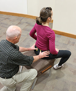
To perform leg fanning, the patient is placed in the seated position toward the front of the palpation stool with the patient's feet 6-8 inches apart. The doctor contacts the medial aspect of each PSIS-sacrum interspace with the thumb pads. Normal motion upon abduction leg fanning is for the sacral base to rotate anterior into nutation and the PSIS to glide down the articulation posterior, inferior and medial.11
 Adduction Leg Fanning
If unilateral fixation is found on abduction leg fanning, the sacrum is relatively posterior and the PSIS is relatively anterior, superior and lateral. According to Cooperstein, the potential listing is AS-lateral ilium on the side of fixation.12
Adduction Leg Fanning
If unilateral fixation is found on abduction leg fanning, the sacrum is relatively posterior and the PSIS is relatively anterior, superior and lateral. According to Cooperstein, the potential listing is AS-lateral ilium on the side of fixation.12
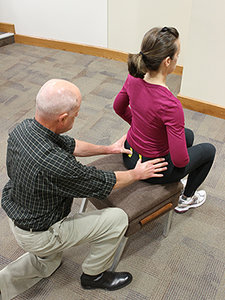
However, normal motion upon adduction leg fanning is for the sacral base to rotate posterior into counternutation and the PSIS to glide up the articulation anterior, superior and lateral.11 If unilateral fixation is found on adduction leg fanning, the sacrum is relatively anterior and the PSIS is relatively posterior, inferior and medial. In this scenario, based on Cooperstein's interpretation, the potential listing is PI-medial ilium on the side of fixation.12
Take-Home Points
As we can see, Cooperstein's "primary listings'"(PI-medial and AS-lateral) are based on the anatomical shape of the sacrum and the oblique glide of the PSIS up and down the articulation upon nutation and counternutation of the sacrum; whereas Gonstead's "married listings'"(PIEX and ASIN) are based on incremental radiographic changes dependent on patient positioning.
Given the evidence of current anatomical and pathophysiological findings, it may be time to re-evaluate the chiropractic hypothesis and our historic understanding of the sacroiliac joint. Recent research is definitely challenging our previous interpretation of the sacroiliac joint and our approach to clinical interventions.
References
- Vleeming A, et al. The sacroiliac joint: an overview of its anatomy, function and potential clinical implications. J Anat, 2012;221:556.
- Vleeming A, et al. Relation between form and function in the sacroiliac joint. Part 1: clinical anatomical aspects. Spine, 1990;15:130-132.
- Egund N, et al. Movements in the sacroiliac joints demonstrated with stereophotogrammetry. Acta Radiol Diagn, 1978;19:833-846.
- Bellamy N, et al. What do we know about the sacroiliac joint? Semin Arthritis Rheum, 1983;12:282-313.
- Bakland O, Hansen JH. The axial sacroiliac joint. Anat Clin, 1984;6:29-36.
- Sturesson B, et al. Movement of the sacroiliac joints: a roentgen stereophotogrammetric analysis. Spine, 1989;14:162-165.
- Vleeming A, et al. Mobility in the sacroiliac joints in the elderly: a kinematic and radiological study. Clin Biomech, 1992;7:170-176.
- Sturesson B. Movement of the sacroiliac joint with special reference to the effect of load. In: Movement, Stability and Lumbopelvic Pain: Integration and Research; Vleeming A, Mooney V, Stoeckart R, editors). Edinburgh: Churchill Livingstone, 2007: pp. 343-352.
- Weisl H. Movements of the sacroiliac joint. Acta Anat, 1955;23:80-91.
- Weinert D. Influence of axial rotation on chiropractic pelvic radiographic analysis. J Manipulative Physiol Ther, 2005;28:117-121.
- Pettersson H. "Leg Fanning." PowerPoint presentation, 2020.
- Cooperstein R. "Because There's a Bone There." J Am Chiro Assoc, Jan-Feb 2013:14-17.
Dr. Howard Pettersson, a 1976 graduate of Logan College of Chiropractic, is an associate professor of technique at Palmer College of Chiropractic. He was the senior editor of Activator Methods Chiropractic Technique – College Edition, published in 1989, and published Pelvic Drop Table Adjusting Technique in 1999. His most recent publication, written with Dr. Green, is How to Find a Subluxation, published in 2003.
Dr. J.R. Green is a 1988 Graduate of Palmer College of Chiropractic. He retired from the Palmer faculty after many years of teaching basic sciences and chiropractic technique. He is currently in private practice in Galva, Ill., and is also an adjunct professor of chemistry with the Eastern Iowa Community College District. Dr. Green was one of the writers of Activator Methods Chiropractic Technique (1997) and also worked as a technical writing consultant on Activator Methods Chiropractic Technique – College Edition and Pelvic Drop Table Adjusting Technique.