


Congenital fusion of cervical vertebrae is a rare anomaly, occurring when two or more cervical vertebrae are congenitally fused. (Fig. 1) This anomaly goes by several names: cervical vertebral fusion syndrome (CVFS), Klippel-Feil syndrome (KFS), congenital dystrophia brevicollis, and Klippel-Feil deformity.1 I consider congenital fusion of cervical vertebrae (CFCV) a better descriptive term, but the literature and most of the research articles refer to Klippel-Feil Syndrome, so to avoid confusion, I will, too.
Key Characteristics
With KFS, there is fusion of two vertebrae which appear structurally as one and function essentially as one motor unit.2 It is rare to have more than two adjacent segments fused together.3 KFS can be distinguished from acquired fused cervical vertebrae such as fusion due to TB, RA or even surgery by two factors: 1) a decrease in the sagittal diameter of the vertebrae, particularly at the site of fusion; and 2) total height of the fused vertebrae is equal to the fused vertebrae's height, including the intervertebral disc.
Additional observations are that the intervertebral discs are absent or there is margin of sclerosis where the "normal" endplates of the segments would have developed. The transverse foramina are smooth; there is a single spinous process for the two vertebral bodies. (Fig. 1)
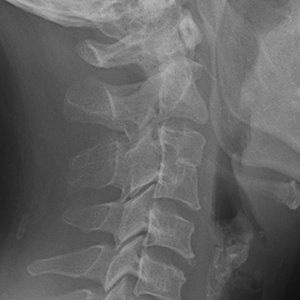 FIG 1 Congenital fusion of C3 and C4.
This anomaly may or may not be symptomatic.4 Often in children there are no symptoms, but with age, even in young adulthood, myelopathy, limitation in neck movement, muscular atrophy and regional sensory loss may develop associated with this anomaly.
FIG 1 Congenital fusion of C3 and C4.
This anomaly may or may not be symptomatic.4 Often in children there are no symptoms, but with age, even in young adulthood, myelopathy, limitation in neck movement, muscular atrophy and regional sensory loss may develop associated with this anomaly.
As chiropractors, we probably see more patients with KFS than anyone in health care because one of the most common symptoms associated with this anomaly is neck pain and stiffness without a history of trauma.
Even though congenital fusion of cervical vertebrae is uncommon, most clinicians are aware of its existence. Some clinicians consider it just a radiographic finding independent from any disease. However, others consider it to be associated with secondary degenerative changes and mobility issues involving adjacent vertebrae.
I believe KFS, even without any associated degenerative changes radiographically, predisposes the patient to biomechanical changes that often lead to early degenerative disc and facet disease.
The most common region of fusion is between the facet joints of the second and third cervical vertebrae (C2 and C3). Clinical signs and symptoms such as shortening of the cervical spine, webbing of the neck, malformations of osseous tissue, limitation of movement, hemi-vertebrae, kyphosis, and lowered line of hair can occur.
Diagnosing this anomaly in childhood can be important in the prevention of secondary degenerative changes in adulthood. Clinicians also must keep in mind that in the presence of vertebral body fusion in the cervical spine, there may be other associated anomalies.
Predisposing Factors / Pathogenesis
A combination of genetic and environmental factors are involved in the pathogenesis of this anomaly: malformation of the notochord, poor performance of retinoids, decreased local blood supply of spine and alterations in genetic expression.5-6
As with most congenital anomalies, the impact of the anomaly will vary from individual to individual.7 With the presence of only one anomaly, the impact will be minimal. But KFS can be associated with other anomalies, such as other vertebral body fusions of other regions in the spine, an abnormal curvature of the spine and/or vertebral instability, spina bifida occulta, raised scapula (Sprengel's deformity), absent rib(s) and other rib defects including cervical ribs, and other skeletal abnormalities.
Even other systems in the body can be affected, including reproductive, urinary, respiratory (breathing problems), cardiovascular, face (cleft palate), ears (hearing difficulties), and eyes.8
KFS can be sporadic, meaning not inherited; or it can be inherited. When it is inherited, it can be inherited as an autosomal dominant or autosomal recessive trait. The disorder begins in the embryotic stage of development when tissue forms into distinct vertebrae.
Researchers are still in the process of teasing out the genetic mutations and variations of KFS presentation.9 For a deeper dive into the phenotypes and categories regarding this anomaly, I refer you to a recent article in Spine.10
Clinical Pearls
The point of this discussion is that patients need to know if they have this anomaly. They should be counseled that they can reduce the development of secondary degenerative disorders by modifying their lifestyle; e.g., avoiding sports that expose them to traumatic injuries to the head and neck, avoiding exercises or activities that involve extensive rotation and extension of the head, etc. These maneuvers may induce compression of the spinal cord and/or the vertebral artery. Even hyperextension of the neck during anesthetic procedures can predispose patients to spinal cord or disc injury.11
When treating patients with this anomaly and other similar osseous congenital anomalies of the cervical spine, clinicians need to be aware of the potential ramifications of this anomaly – not only chiropractors, but also spine surgeons, physical therapists and even the anesthetist when performing procedures, such as endotracheal intubation.
These patients can respond well to chiropractic, but the clinician needs to be aware of the levels of fusion to avoid encouraging hypermobility at the adjacent mobile segments. There are many techniques a chiropractor can use to manage these patients; one that has been well-documented in the literature is the "flexion distraction technic" developed by Dr. James Cox.12
References
- Dunsker SB, et al. Craniovertebral anomalies. Clin Neurosurg, 1980;27:430-9.
- Erdil H, et al. Congenital fusion of cervical vertebrae and its clinical significance. J Anat Soc India, 2003;52:125-127.
- Brown MW, et al. The incidence of acquired and congenital fusions in the cervical spine. Am J Roentgenol Radium Ther Nucl Med, 1964;92:1255-1259.
- Singh A, et al. Congenital fusion of typical cervical vertebrae. MOJ Anat Physiol, 2016;2:166.
- Sadler TW. Langman's Medical Embryology, 6th Edition. Baltimore: Williams and Wilkins, 1990: p.p. 151-153.
- de Graaff R. Congenital block vertebrae C2-3 in patients with cervical myelopathy. Acta Neurochir (Wien), 1982;61:111-126.
- Paraskevas GK, et al. Congenital synostosis of cervical vertebrae: an osteological study and review of the literature. Cureus, 28 Oct 2019;11(10):e605.
- Mardani M, et al. Congenital fusion of cervical vertebrae: a review on embryological etiology. Rev Clin Med, 2016;3(4):148-153.
- Klippel Feil Syndrome. Genetic and Rare Diseases Information Center (website). https://rarediseases.info.nih.gov/diseases/10280/klippel-feil-syndrome
- Hachem LD. et al. Klippel Feil Syndrome: clinical phenotypes associated with surgical treatment Spine, 2020;45(11):718-726.
- Soni P, et al. Cervical vertebrae anomalies-incidental findings on lateral cephalograms. Angle Orthod, 2008;78:176-180.
- Kruse R, Cox J. Case report: treatment of adjacent segment disease related to a C5-C6 block vertebra (Klippel-Feil Disease). ACA News, October 2017.
Click here for more information about Deborah Pate, DC, DACBR.