


Review the cervical flexion radiograph in Figure 1A. The patient has an increased atlantodental interval (ADI) at C1-C2. Now review the cervical extension radiograph of the same patient in Figure 1B. The ADI decreased. Why?
To be thorough, other instabilities in the upper cervical region of Down's patients must be mentioned. They include instabilities that result from atlas and axis deformities and atlanto-occipital deformities. However, the ADI is our focus here.
ADI Instability: Signs and Potential Spinal Consequences
Down's syndrome is a congenital reason for an unstable ADI. However, there are also pathological reasons. Rheumatoid arthritis commonly affects the atlantodental articulation by eroding the transverse ligament, and trauma can rupture the ligament.
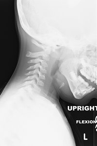 Figure 1A
The primary concerns for a patient with an ADI instability are additional undo stresses placed on the already unstable area and the spinal cord. When the head moves forward, the atlas moves with it. If the head is moved rapidly, such as in trauma, the head's weight can cause further ligamentous damage. As the atlas moves forward, its posterior arch places pressure on the posterior aspect of the spinal cord, creating a shearing force and displacing the cord anteriorly.
Figure 1A
The primary concerns for a patient with an ADI instability are additional undo stresses placed on the already unstable area and the spinal cord. When the head moves forward, the atlas moves with it. If the head is moved rapidly, such as in trauma, the head's weight can cause further ligamentous damage. As the atlas moves forward, its posterior arch places pressure on the posterior aspect of the spinal cord, creating a shearing force and displacing the cord anteriorly.
In atlantodental instability, it is not unusual for the patient to report that he/she feels the shift of the unstable segment when changing from one posture to another. This information is a crucial clue to instability. The sensation will be described as a "deep clunk," which differs from the sensation associated with typical joint cavitation.
Imaging Clues to Look For
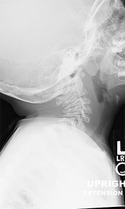 Figure 1B
During the initial patient examination, if imaging is performed, usually the traditional AP, APOM and lateral view series, instability may or may not be identified. An increased ADI can be observed on a neutral lateral view, but not always.
Figure 1B
During the initial patient examination, if imaging is performed, usually the traditional AP, APOM and lateral view series, instability may or may not be identified. An increased ADI can be observed on a neutral lateral view, but not always.
For this reason, the traditional three-view series plus flexion and extension views are recommended for patients with Down's, rheumatoid arthritis, or significant head and neck trauma.
Key Orthopedic Tests
Another option for identifying instabilities, one usually performed before imaging, is orthopedic testing. While less accurate, orthopedic testing is fast, adds minimal time to the course of an examination, and does not involve radiation and additional expense.
These tests are performed for the same reasons the traditional three-view series plus flexion and extension are performed for patients with Down's, RA, or significant head and neck trauma.
Accuracy is of interest because instabilities other than that caused by transverse ligament absence or pathology can be present, increasing the possibilities in the differential diagnosis. (Note those mentioned above in the discussion of Down's syndrome.) Four orthopedic tests are recommended:
- Lateral displacement test
- Aspinall's test
- Upper cervical flexion test
- Sharp-Purser test
How to Perform the Tests
The first three tests are all performed with the patient in a supine position. The examiner is at the head of the exam table. The Sharp-Purser test is performed with the patient supine, but the examiner is positioned to the side of the table.
The lateral displacement test assesses the integrity of the transverse ligament. The examiner contacts the lateral aspect of the atlas on one side and the lateral aspect of the axis on the opposite side. A lateral-to-medial force is applied at each contact point.
The examiner feels for side-to-side movement between the atlas and axis. The examiner then repositions his / her hands to the atlas and axis on opposite sides. The lateral-to-medial forces are reapplied. Detection of side-to-side movement between the atlas and axis remains the abnormal finding.
Aspinall's test assesses the integrity of the transverse ligament. The examiner contacts the occiput and atlas firmly to stabilize the contact between the two structures. The examiner then flexes the cervical spine and holds the position. Finally, a posterior-to-anterior force is applied to the posterior arch of the atlas.
The test is positive if the patient/examiner feels a shift of the atlas. The patient might also describe the sensation of a lump in his / her throat. The sensation is said to be created by the atlas moving forward. The shift may be associated with an audible "clunk."
The upper cervical flexion test assesses the integrity of the transverse ligament. The examiner contacts the posterior arch of the atlas with one hand while contacting the occiput with the other. The examiner then places his / her shoulder against the patient's forehead and induces a slight ventral flexion at the contact areas. Excessive movement and possibly a "clunk" may be produced, indicating ADI instability.
The Sharp-Purser test assesses the integrity of the transverse ligament. The examiner uses his / her thumb and index finger to pinch the spinous process of the axis; and then places pressure in an anterior-to-posterior direction on the patient's forehead.
If the patient and doctor feel the head shift in a posterior direction, the test is positive for the ADI reduction. The shift may be associated with an audible "clunk."
The first three tests are intended to increase the ADI and recreate symptoms. Sharp-Purser is intended to decrease the ADI and decrease symptoms.
Clinical Takeaway
The importance of this topic relates to clinical safety. Manipulation of an unstable area is contraindicated. Referral for a second opinion is necessary, as the ADI may require surgical stabilization.
Adjusting other areas of the patient's spine is reasonable if those areas are stable. The patient can still benefit from chiropractic care with caution employed.
Author's Note: Radiographs courtesy of Julia Crim, MD, University of Missouri School of Medicine.
General Resources
- Yochum T, Rowe L. Essentials of Skeletal Radiology, Volume 1, 3rd Edition. Lippincott, Williams and Wilkins, 2005.
- CORE, Clinical Orthopaedic Exam: A Diagnostic Utility and Reference Tool for Musculoskeletal Disorders. Clinically Relevant Technologies, 2010.
- Magee DJ. Orthopaedic Physical Assessment, 5th Edition. Saunders-Elsevier, 2008.
Click here for more information about K. Jeffrey Miller, DC, MBA.