


The lateral thoracic view can be challenging because to visualize the cervicothoracic junction often requires an additional view. In fact, I find that this cervicothoracic junction is perhaps the most difficult area to demonstrate on plain films.
The true difficulty with this view is knowing what's going to work with which patient. Let's review the most common techniques for obtaining a swimmer's lateral view of the cervicothoracic junction. None of these techniques will work all of the time, but it helps to know about each of them so you can, at the very least, make an educated guess as to what might work with a particular patient. Keep in mind that this view has the worst of everything: high dose, high scatter, difficult positioning, doesn't work well on large patients or patients with shoulder problems; and to boot, it has some critical anatomy.
Arms Forward
The humeral heads are the problem with all of these positions. The whole idea is to try and get the humeral heads projected away from the cervicothoracic junction. The arms-forward technique involves moving both humeral heads anteriorly by having the patient cross their arms in front of them; or by extending their arms out in front of them by holding onto a bar attached to the upright bucky. This technique usually works if the patient's shoulders are flexible. As with all lateral thoracic techniques, using a breathing technique will help blur the lung markings.
One Arm Up, One Arm Down
This is the classic swimmer's technique. This version of the technique achieves an unobscured lateral view of the cervicothoracic junction by positioning one arm up and one arm down. Anatomically, it doesn't matter which arm is up and which is down. One of the flaws of this technique is that you tend to laterally flex the patient. This causes a loss of visualization of the joint spaces.
Pulling the Arms Down
This technique is often utilized when you have come close to demonstrating the segment of interest, but not quite. The reasoning is that by moving the shoulders inferiorly (and with a little bit of luck), you will achieve a clear view of the segment in question. The way to achieve this slight lowering of the shoulders is to give the patient a rope to stand on so they can pull their own arms down. Of course, if this maneuver causes any symptoms it should be discontinued.
Supine Lateral (Modified Swimmer's)
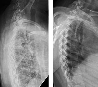 Left: Lateral thoracic with no breathing technique; Right: Lateral thoracic with breathing technique.
This technique can be used in patients who do not have an acute injury and can lie on their side. The position is very similar to a lateral thoracic spine position, except the arm that is away from the table is rolled posteriorly. Obviously this requires some flexibility by the patient. The disadvantage of this technique is that it is an off-lateral position, but it might be a better trade-off than not being able to visualize the anatomy.
Left: Lateral thoracic with no breathing technique; Right: Lateral thoracic with breathing technique.
This technique can be used in patients who do not have an acute injury and can lie on their side. The position is very similar to a lateral thoracic spine position, except the arm that is away from the table is rolled posteriorly. Obviously this requires some flexibility by the patient. The disadvantage of this technique is that it is an off-lateral position, but it might be a better trade-off than not being able to visualize the anatomy.
Breathing Technique
All of these techniques can be greatly improved with the use of a breathing technique. (Figure 1A and 1B) Remember that an exposure time of 1 second or greater is required to achieve sufficient blurring of the thoracic soft tissues. You will have to adjust the technical factors accordingly.
These are the most commonly used techniques to evaluate the cervicothoracic junction with plain films. Of course, with the ability to use CT scans, this area is not nearly as difficult to image, although increased exposure to radiation is something we always need to consider.
Click here for more information about Deborah Pate, DC, DACBR.