Hip pain is a term that is used loosely by patients. When patients tell me their hip hurts, I ask them to point at the perceived source of the pain. They will usually point at the sacroiliac joint area, the gluteal muscles, or the greater trochanter area. When the trochanter is tender, the medical diagnosis is usually trochanteric bursitis. I consider this a "garbage can" diagnosis - naming a syndrome for the location of the pain, and then treating the local pain with steroid injections, rather than finding the underlying combination of dysfunctions that will solve the problem.
How do I determine that the hip, meaning the junction of the femur and the acetabulum, is part of the problem? I'll use my usual tools: listening, palpating, and therapy localizing with indicator muscle testing. Range-of-motion testing is also very valuable.
For the hip joint itself, two main joint restrictions seem to be critical. The first is an anterior femur, whereby the ball of the femoral head is functionally anterior, and resists posterior glide. Look for this pattern in patients with knee pain. I have some skepticism about reflex-based muscle testing that specifies that a certain muscle weakness indicates a particular lesion, but I have found that an anterior femur is almost always accompanied by weakness of the hip flexors. I test this by having the patient supine, with the whole leg flexed anterior at 20 degrees, and seeing if they can hold the leg up while I push straight posterior. This tests more the long hip flexor, the rectus femoris, rather than the psoas. I suspect that this weakness is caused by the mechanical disadvantage created by the anterior hip subluxation pattern.
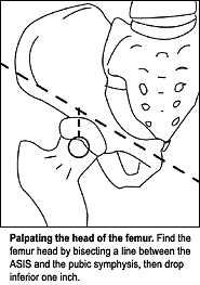
The anterior femur has a clear tenderness indicator: an exquisite tenderness directly over the head of the femur. The image below illustrates this location. You can palpate the head of the femur by bisecting the inguinal ligament, halfway between the ASIS and the pubes, and then moving your thumb inferior about 1 centimeter. You'll be over an obviously bony area. When the head of the femur is stuck anterior, this bony prominence will be more palpable than usual, as well as tender. Compare to the other side the first several times you assess here, until you get a clear sensory "picture" of the feeling of this subluxation. Remember that you are dealing with a sensitive area, in the groin, so use the usual precautions for potentially sexually charged areas. I always tell the patient what I am doing, make eye contact, and explicitly ask permission to assess and work in this area.
The correction here is quite simple. The easiest way to correct is recoil (engage-release). You do not need to use significant force. Engage the restriction in three dimensions, starting with anterior to posterior pressure, and add pressure from either the medial or lateral side, whichever feels more restricted. Finally, add a torquing force, either clockwise or counterclockwise. I am using the words "pressure" and "torque," but I am always extremely gentle. Remember to keep your hands in neutral, and not bias the assessment via your preloading direction. If you continually find the exact same direction, you are probably fooling yourself. Engage to the "feather edge" of the barrier, then suddenly release outward. You can use the patient's respiration to further enhance the adjustment, doing the adjustment on the phase of the breath that increases the feeling of resistance. I never do a thrust type of adjustment here, and rarely use Engage Listen Follow.
The second major hip joint restriction is when the hip is stuck in external rotation, or resists internal rotation. This is consistent with the capsular pattern of the hip, where the first movements that are lost in hip problems are flexion and internal rotation. You can test these through gross range of motion testing, looking for a lack of motion in both flexion and internal rotation and testing for end feel. Remember that on passive hip flexion, the hip motion ends as soon as pelvic motion begins. The patient who has lost the internal rotation hip motion has a hip that is headed for trouble. If this motion does not return with your manipulation and soft-tissue work, the hip may be already severely degenerated. Conversely, keeping this internal rotation motion functional may help the patient put off hip replacement surgery for years. When the hip is stuck in external rotation, the foot will "flop" out laterally as the patient lies flat. The externally rotated hip will affect the whole lower extremity chain, putting a valgus stress on the knee, and increasing pronation in the foot. We need to address these issues from below, with orthotics and adjusting of the foot, and from above, by normalizing hip motion.
I assess hip internal rotation with the patient supine, and with the thigh bent up to 90 degrees if possible, and the knee bent at 90 degrees. My proximal hand stabilizes at the knee, and my lower hand uses the ankle as a lever to pull laterally, torquing the hip internally. Normal motion on a middle-age male is about 15 degrees; females often have significantly more motion. You can compare to the other side. You are also looking at the end feel of the joint; a hard end feel with minimal motion indicates a need for the adjustment here.
The adjustment is quite simple. I use a quick thrust here. It's not really a "low-force" adjustment in the same sense that most of the Framework techniques are. It's a classic high-velocity, low-amplitude chiropractic thrust. It doesn't take much force, just quickness.
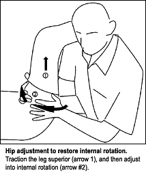
I'll use the right leg for our example, as illustrated in the picture [on page 48]. The patient is supine, with the right leg in the same 90-90 position I described above. I am sitting on the patient's right side, with my right shoulder against their posterior thigh. I use both of my hands to grasp firmly around the whole of the upper thigh. My left hand will be the main hand, with the heel of my left hand pushing against the greater trochanter. I pre-tension the area by both lifting the whole thigh superior and taking it into internal rotation to the feather edge of the barrier. I then do a quick thrust into further internal rotation. I really don't use much fine-tuning of three dimensions here; it's a pure thrust into internal rotation.
The results of this adjustment are often dramatic and immediately visible. When it's right, you will instantly get much more hip internal rotation. Do your post-check; it will usually be very satisfying for both you and your patient after this adjustment.
The key rehab exercises for an externally rotated hip involve stretching of the piriformis and strengthening the gluteus maximus and medius, as discussed in several previous articles. In an anterior hip, the quadriceps will probably have weakened to some degree, so terminal knee extension and modified squats and lunges may be beneficial.
The major soft-tissue area to look at here, besides the piriformis and gluts, is the quadratus femoris. This little muscle goes from the ischial tuberosity laterally to the posterior side of the greater trochanter. It will often be tight and tender, especially at its insertion into the trochanter when the hip isn't internally rotating. Use your favorite soft-tissue methods to release it.
The hip is a key link from the pelvis to the lower extremity. Correcting problems here can help pain and dysfunction up and down the kinetic chain.
References and Resources
- Thomas, Mark DC. Extremity adjusting classes, 01-03.
- Cyriax, James. Textbook of Orthopedic Medicine: Diagnosis of Soft Tissue Lesions, Vol. 1; 1975.
- Chauffour, Paul. Mechanical link courses, 1997-2001.
Click here for more information about Marc Heller, DC.


