


I have personally used this diagnostic procedure in acupuncture since 1973. Prior to that time, I practiced pulse and tongue diagnosis. Even though I was obviously a neophyte in 1973, electro-meridian imaging (EMI) allowed me to practice at the master level. (As pointed out in my previous article, I renamed the ryodoraku procedure to EMI in 1982.)
In my previous article, I illustrated the procedure of EMI by detailing a diagnostic graph on a pre-printed form that could be obtained without the benefit of a computer by simply adding all of the measurement figures of the source points together, then dividing by 24 to develop a base average. By establishing the normal range for each patient based on that formulation, the graph clearly shows which meridians are energetically involved.
However, one of the most exciting applications of EMI is the computer-enhanced version. With a computer, the procedure takes less than two minutes to perform. Clinical response using this diagnostic procedure is legendary. This article will focus on several case histories from my personal patient files illustrating the graphing from the computer-enhanced EMI. They are typical examples of the significance of this procedure. This method of diagnosis can potentially be an incredible addition to your practice regardless of your style of acupuncture.
Case History #1
The patient, a 27-year-old male, fills out the preliminary medical history and other entry forms. During the consultation, I discover the patient has been referred to me for severe adult onset asthma. His situation is extremely grave. He has been taken to the hospital three separate times by ambulance. The patient had no history of asthma or respiratory distress until the sudden onset of asthma four years ago. He has seen numerous physicians. He reports extreme shortness of breath in the middle of the night, specifically between the hours of 3 and 5 a.m., which is consistent with the horary cycle in the midday-midnight law of acupuncture.
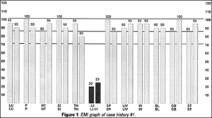
EMI examination reveals that all meridians are normal except the large intestine, which is pathologically depressed (see Figure 1). Upon further questioning, the patient asks if the low large intestine meridian reading could be a result of his colostomy. Colostomy? There was no mention of a colostomy in his medical history form. The patient explains he was on a church mission in the Australian outback and developed severe intestinal distress from contaminated water, which led to the surgical removal of his large intestine and half of his small intestine. I then learned his asthma developed within the first two weeks following his colostomy surgery.
Based on the acupuncture principles, lung is paired with large intestine, and what affects one could affect the other. It was reasoned his asthma may be caused by his extremely depressed large intestine meridian.
The treatment was elementary. I used the tonification point for the large intestine meridian (LI 11), followed by the mu (alarm) point (ST 25) and the shu (associated) point (BL25). No treatment was geared for his lung as either an organ or meridian. The entire treatment revolved simply around his large intestine meridian as discovered by the EMI examination.
The patient was given two treatments, followed by a new EMI graphing. The new EMI showed a correction of the problem. The patient remained a patient, receiving maintenance-type treatments four times a year with routine EMI examinations twice a year. He remained a patient for eight years, then relocated to another city, at which point I lost track of him. In the eight years of follow-up care, he never experienced another asthma attack or even shortness of breath. He was virtually asymptomatic following the second treatment.
Case History #2
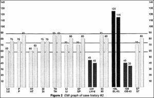
A 65-year-old female was referred by a physician in another state in the hope that acupuncture treatment would bring about a clinical response. She has extreme femoral nerve neuritis of three years' duration, and is unable to walk more than 7-10 steps without severe, disabling pain. The patient has had extensive physiotherapy, and six months of chiropractic care. She's been under the care of a neurologist and an orthopedist, who prescribed a variety of medications that the patient states has had little if any effect.
During the EMI examination, it was learned that she had gallbladder surgery just prior to the onset of the condition. She returned for a second surgery to surgically remove scar tissue that had apparently formed as a result of the first surgery. The patient was treated according to the protocols set forth in the EMI examination to tonify the liver (L8), to sedate the bladder (BL65), and to tonify the gallbladder (GB 43). She further was treated with laser beam stimulation directly to the abdominal scar tissue. Her debilitating pain was completely and permanently resolved. She received two treatments from me and was referred to her local practitioner for follow-up care.
Every Christmas I receive a special card from her thanking me for acupuncture.
Case Histories #3 and #4
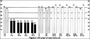
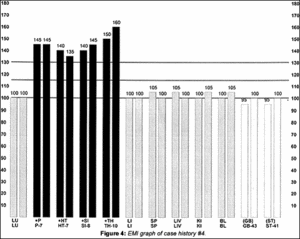
These two case histories share the identical symptomatology of severe panic attacks. Both cases share common findings, making their conditions seem almost exactly the same. They both have dramatic stories of disabling panic attacks. Neither of them can leave the house to perform simple tasks like running to the grocery store or picking up the kids after school without grave consequences. They are both confined to their homes. The sheer thought of going out of the house is overwhelming.
Even though their symptoms are practically identical, the EMI graphs are polar opposites. One graph shows a very depressed pericardium, heart, small intestine and triple heater meridian; the other shows each of those same meridians to be elevated. The rule in EMI evaluation is likened to, "This bed was too hard, this bed was too soft, but this one is just right." It makes little difference if the meridians are too high or too low. If the meridians are involved, it is clinically significant. Just as one may tune the radio to 94.5 for a clear station, 94.6 and 94.4 will broadcast static. There is nothing wrong with the radio, just something wrong with the fine-tuning. The meridians comprising the fire element are involved. This is a classic finding for panic attacks. This condition is very responsive to acupuncture when balanced. Both of the patients whose graphs are shown had incredible responses.
I have thousands of interesting cases in my patient files I would love to share with you, however, as you are aware, space in this publication is at a premium. For further information on EMI, drop me an email at
![]() . Best wishes for a fantastic holiday season!
. Best wishes for a fantastic holiday season!
John Amaro,DC,FIAMA,Dipl.Ac.,LAc
Carefree, Arizona
Click here for previous articles by John Amaro, LAc, DC, Dipl. Ac.(NCCAOM), Dipl.Med.Ac.(IAMA).