


Many of the ligamentous (and tendon origin) and insertions of the muscles and ligaments around the pelvis can appear very unusual and yet be just degenerative changes. They appear like bony proliferations or areas of rarefaction, or even areas of sclerosis or ossification. The key factors are that these changes are not associated with those in the underlining bone and they are exacting where the tendon or ligament attaches to it.
The diagram below demonstrates the most common sites in the pelvis where these areas of enthesopathy and rarefaction generally are seen. The sites of predilection for fibro-ostoeoses (left, shaded black, sharply delineated) and fibro-osteitis (right, shaded gray) are often associated with increased density of surrounding tissues in the region of pelvis and hip. Ligamentous ossifications (from above downward = iliolumbar, sacrospinal, tuberosacral, pectineal ligament and ligamentum superius pubis) indicated by dashed lines.
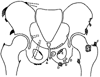
Figure 1: Fibro-ostosis/Fibro-osteitis
Specific sites:
A= fibro-osteosis of muscularis gracilis
B= rarefying fibro-osteitis of the arcuated pubic ligament
C= rarefying fibro-osteitis at the origin of the adductor longus and brevis muscles
D= productive and rarefying types of fibro-osteitis in the ischium
E= fibro-osteitis at the origin and insertion of the obturator externus muscle
F= rarefying and productive fibro-osteitis at the insertion of the gluteus medius
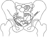
Figure 2
Of, course there are certain diseases that are also associated with these same proliferative changes, such as ankylosing spondylitis, hyperparathroidism, fluorosis, ochronosis, excessive occupational strain, and trauma. But most of the time the cause of these radiographic findings are due to conditions such as degenerative joints disease (DJD) and diffuse idiopathic hyperostosis (DISH).
The following diagram (see Figure 2) pertains to benign findings in the pelvis. There are many shadows that appear on the AP pelvis film that are generally not of great concern. They are visible only on occasion. It depends on the patient's individual habitus and the x-ray technique employed.
This diagram is of the most common pelvic soft tissue shadows that can be seen on the AP pelvis view. The most frequently recognizable are the bladder filled with urine and gas in the loops of bowel.
Diagrams and reference information: Wolfgang Dihlmann's Joints and Vertebral Connections: Clinical Radiology, Thieme Inc. 1985.
Deborah Pate,DC,DACBR
San Diego, California
![]()
Click here for more information about Deborah Pate, DC, DACBR.