


The simple bone cyst, also referred to as a unicameral or solitary bone cyst, is a common nonneoplastic lesion of unknown origin. Originally, this lesion was thought to be a tumor, a post-infectious cyst or possibly a dysplastic region of bone.1-3 In the late 1960s to early 1970s, the fluid contained within the simple bone cyst was analyzed and shown to be consistent with serum. In addition, it was discovered that there is stasis of the fluid within the lesion upon injection of contrast material.1-3 Since that time, the simple bone cyst has been considered to be the result of altered venous drainage and accumulation of interstitial fluid in a rapidly growing region of cancellous bone. 1-3 It is still categorized as a tumor and represents approximately 3 percent of biopsied primary bone tumors.2
Who Gets It?
The simple bone cyst is seen more often in males with a ratio of 2-3:1 over females. It is seen primarily in the younger population below the age of 20. Approximately 80 percent of the cases occur between the ages of 3-14, with the average age being 9.1-3 There have been documented cases occurring as early as two months and others seen as late as 50 years of age.2 By far, male patients younger than age 20 should be considered most likely to suffer a simple bone cyst.
Where Does It Occur?
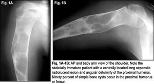 In approximately 90 percent to 95 percent of cases, a simple bone cyst will occur in the long bones, with 56 percent frequency in the humerus, 27 percent frequency in the femur, and 6 percent in the tibia, among reported cases.1-3 In patients older than age 20, there is a higher rate of occurrence in the flat or irregular bones.1 Exceptions are the calcaneus and the small bones of the hands and feet, which rarely are affected.1 In addition, there are rare cases reported in the ischium, sacrum, pubic bone, ribs, patella, scapula, clavicle and spine.1,2 By far, the most common location is the proximal humerus, and rarely will there be more than one lesion encountered in a single patient.1-3
What Does It Look Like?
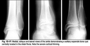
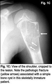 The classic radiographic appearance of the simple bone cyst is a centrally located, radiolucent lesion that thins the cortex of surrounding bone due to expansion.1-3 (See Fig. 1A-1F.) It most frequently presents as a single chamber (unicameral) but can be multilocular. In either appearance, there is a well-defined border and sclerotic edge. The result is a geographic pattern of lucency with a short zone of transition between abnormal and normal bone.1-3 Since the lesion is not painful, it may be present for years. As the patient grows, the lesion will elongate with the long axis of the affected bone and migrate distally toward the diaphysis. It is common for the length of the cyst to be greater than the width.1,2 There is no associated soft tissue mass or periosteal reactions, although they may be present in the event of a fracture.2 In about 10 percent of cases with a fracture, there is a finding called the "fallen fragment sign." This refers to a detached fragment of bone that is seen floating within the cyst.2 Based on the plain-film findings, other lesions to be considered in a differential diagnosis include aneurysmal bone cyst, fibrous dysplasia or enchondroma.1
How Do We Treat It?
This lesion is not painful and often is detected as an incidental finding. In approximately 65 percent of patients, a bone cyst is found as the underlying pathology associated with a fracture.3 Treatment consists of curettage and grafting, excision and more recently, a common procedure consisting of percutaneous steroid injection.1-3 Recurrence rate of the lesion after curettage is approximately 20 percent.3 The main reason to address this lesion is to prevent pathologic fracture and deformity.1-3
 Conclusion
Although not a tumor, the simple bone cyst has remained in this category since its discovery in the 1940s. The patient typically is a young male below the age of 20 with an expansile cyst occurring in the proximal humerus or femur (90 percent of the time).
The common clinical presentation is that which is associated with a pathologic fracture; management consists of an orthopedic referral. The patient will receive either steroid injection or possibly surgery. Co-management and rehabilitation are appropriate for the treating chiropractor.
References
- Resnick D, Niwayama G. Diagnosis of Bone and Joint Disorders, 3rd ed. Philadelphia: W.B. Saunders 1995.
- Yochum T, Rowe L. Essentials of Skeletal Radiology, 3rd edition, Baltimore, Williams & Wilkins 2005.
- Marchiori D. Clinical Imaging with Skeletal, Chest and Abdomen. St. Louis, Mosby 1999.