Twelve percent of the U.S. population ages 25 to 75 years has symptoms and signs of osteoarthritis (OA). There are three major risk factors associated with the development of OA: body-mass index, trauma and heredity.
Modifiable Risk Factors
BMI is by far the major factor in the disease process. Obesity increases the overall loading of the knee and has an accelerating effect on the progression of the disease. Malalignment of the knee is also a factor in the progression of OA.1 Studies indicate that varus, valgus, and leg-length discrepancy contribute to the progression of the disease. Although elevated BMI increases the risk of knee OA progression, the effect of BMI is limited to knees in which malalignment exists. This is because of the combined focus of load from malalignment and the excess load from increased weight. BMI and malalignment are factors that can be managed. Both should be addressed if conservative management is to be successful.
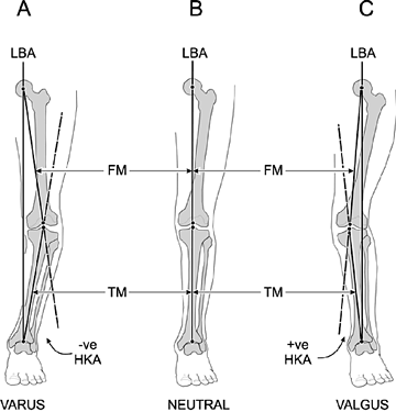
At the knee, alignment (i.e., the hip-knee-ankle angle) is a key determinant of load distribution. Any shift from a neutral alignment of the hip, knee and ankle affects load distribution at the knee. The most common method of evaluating alignment of the knee is to draw a line from the mid-femoral head to the mid-ankle. This line represents the load-bearing axis.
In a varus knee, this line passes medial to the knee and a moment arm is created, which increases force across the medial compartment of the knee. In a valgus knee, the load-bearing axis passes lateral to the knee, and the resulting moment arm increases force across the lateral compartment of the knee. Statistical studies indicate there is not much room for error as far as alignment goes; deviation of more than 1.3 degrees from neutral becomes a risk factor for OA.1
This method, however, may not always be practical; an alternative method is to take a single AP view of the knee and measure deviation of the center points of the femoral condyles, center of the tibial spines and the center of the tibial plateau. (Visit www.ncbi.nlm.nih.gov/pmc/articles/PMC2663322/figure/fig1/ to view an illustration of the method of calculating the local tibiofemoral alignment angle on a short anteroposterior knee radiograph.) This method, described by Kuhn, et al.,2 in their article, "Effect of Local Alignment on Compartmental Patterns of Knee Osteoarthritis," is a more practical method and has been demonstrated to be reliably accurate.
The other alignment risk factor is leg-length discrepancy, which can be determined with the classic method of a scanogram3 or with digital CT, which offers less radiation exposure.4 Generally, a threshold difference of 10-12 mm is considered clinically meaningful. A fairly recent study5 performed with the MOST data6 determined that a difference of 1 cm becomes a risk factor for OA on the short-leg side. This is very interesting since an estimated 50-60 percent of the population has a leg-length discrepancy.
With the combination of risk factors including obesity, malalignment and leg-length discrepancy, a large sector of the population is at risk of developing OA before the age of 70; often decades earlier. Current national estimates are that by the year 2020 18.2 percent of the U.S. population (59.4 million) will be affected by OA. These statistics underscore the need to develop effective interventions that reduce the stresses imposed by a given malalignment.
Conservative Treatment
We don't have any medical treatment to date that will prevent OA, nor do we even have any medical treatment that will slow its progression. What we do know is that obesity, knee alignment and leg-length discrepancy are factors that affect the incidence and progression of OA, and these factors can be managed.
Most chiropractors are treating knee malalignment in their patients as a matter of course because of their concern for structural alignment. More than any other health care professional, we understand alignment. Besides the obvious, which is getting patients on a weight loss program, we need to address the alignment issue. With proper use of orthotics and specific exercises, the alignment factor can be managed conservatively, especially in cases of mild to moderate OA.
For example, Kuhn, et al.,7 have documented significant improvement in the Q-angle in hyperpronating male subjects with the use of flexible orthotics. Immediate changes in the quadriceps femoris angle were noted after insertion of an orthotic device. Saxena, et al.,8 concluded that semiflexible orthoses showed a significant decrease in the level of pain in patients with patellofemoral pain syndrome. Eng, et al.,9 came to same conclusion that orthotics relieve symptoms associated with patellofemoral pain syndromes.
Just recently (Dec. 16, 2010 issue), Dynamic Chiropractic published an article by Mark Charrette, DC, titled "When to Consider Orthotics: Research-Based Recommendations,"10 in which Charrette reviews the clinical and diagnostic findings indicating when orthotics should be considered.
 VARUS / VALGUS ALIGNMENT OF THE KNEE
LBA: load-bearing axis; HKA: hip-knee-ankle angle; FM: femoral mechanical axis; TM: tibial mechanical axis.
A. Varus: center of the knee is lateral to the LBA line and the HKA angle is negative.
B. Neutral alignment: LBA line and HKA angle are colinear.
C. Valgus: center of knee is medial to the LBA and the HKA angle is positive.
Exercise is the last piece of the treatment program. It is well-documented that resistance training improves muscle strength and endurance. Resistance training has also been shown to improve function of patients with knee OA.11-12 In general, strengthening the quadriceps has been a common goal in the management of knee OA. In healthy knees, strength protects against the development of OA; in arthritic knees, however, greater strength does not always translate into improved function if the alignment is not addressed. Sharma, et al.,13 demonstrated that malalignment or laxity and greater strength may actually damage the joint increasing the progression of the arthritic changes.
Specifically designed exercises and orthotics are of major importance when managing patients with knee OA. We can't just send patients to the gym with a list of generic exercises. Alignment has to be addressed first; once that is improved, specific exercises for the realignment and strengthening of the lower extremity can be incorporated.
It is finally being recognized that joint alignment is a major factor in the development and progression of OA. Chiropractors have been managing patients with OA for decades with the idea of improving joint alignment. Much of the chronic problems associated with OA can be improved with chiropractic management.
References
- Sharma L, Song J, Felson DT, et al. The role of knee alignment in disease progression and functional decline in knee osteoarthritis. JAMA, Aug. 15, 2001;286(7):
- Khan FA, Koff MF, Noiseux NO, et al. Effect of local alignment on compartmental patterns of knee osteoarthritis. J Bone Joint Surg Am, Sept. 1, 2008;90(9):1961-1969.
- Sabharwal S, Zhao C, McKeon JJ, et al. Computed radiographic measurement of limb-length discrepancy: full-length standing anteroposterior radiograph compared with scanogram. J Bone Joint Surg Am, 2006(88): 2243-2251.
- Sabharwal S, Kumar A. Advances in limb lengthening and reconstruction. Clin Ortho & Rel Res, 2008; 466(12): 2910-2922.
- Harvey WF, Yang M, Cooke TDV, et al. Association of leg-length inequality with knee osteoarthritis: a cohort study. Annals Int Med, March 2, 2010;152(5).
- Funded by the National Institutes of Health, the MOST is a multicenter, longitudinal, community based study of 3,026 participants ages 50 to 79 years with knee osteoarthritis or with high risk for knee osteoarthritis due to knee pain, obesity, knee injury or knee surgery.
- Kuhn DR, Yochum TR, Cherry AR, et al. Immediate changes in the quadriceps femoris angle after insertion of an orthotic device. J Manipulative Physiol Ther, September 2002;25(7): 465-470.
- Saxena A, Haddad J. The effect of foot orthoses on patellofemoral pain syndrome. J Am Podiatr Med Assoc, 2003 Jul-Aug;93(4):264-71.
- Eng JJ, Pierrynowski MR. Evaluation of soft foot orthotics in the treatment of patellofemoral pain syndrome. Phys Ther, February 1993;73(2):62-8; discussion 68-70.
- Charrette M. "When to Consider Orthotics: Research-Based Recommendations." Dynamic Chiropractic, Dec. 16, 2010.
- Farr JN, Going SB, McKnight PE, et al. Progressive resistance training improves overall physical activity levels in patients with early osteoarthritis of the knee: a randomized controlled trial. Physical Therapy, March 2010;90(3).
- Baker KR, Nelson ME, Felson DT, et al. The efficacy of home based progressive strength training in older adults with knee osteoarthritis: a randomized controlled trial. J Manipulative Physiol Ther, Sep;25(7):465-70
- Sharma L, Dunlop DD, Cahue S, et al. Quadriceps strength and osteoarthritis progression in malaligned and lax knees. Ann Intern Med, April 15, 2003;138:613-619.
Click here for more information about Deborah Pate, DC, DACBR.


