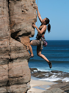
Anterior elbow pain is commonly associated with problems of the musculotendinous complex of the biceps muscle. However, anterior elbow pain can also be associated with a lesser known musculotendinous disorder involving the brachialis muscle: climber's elbow.1
The recent trend of cross fitness training has the potential to add to the number of brachialis injuries. Pull-ups, chin-ups and rope climbing frequently performed in cross fitness programs have been known to cause brachialis injury at the elbow. With the availability of cross fitness programs in gyms and training facilities, cross fitness participants will soon outnumber those participating in rock climbing. This means doctors practicing in areas where climbing sports are not common may begin to see more brachialis injuries.
Diagnosing the Condition
 Diagnosis of this condition begins with the patient's history. Noting specific signs and symptoms and participation in any of the activities listed above is important.
Ruling out the presence of the more common bicipital injury at the elbow is the next logical step. The distal biceps and/or its tendon should be palpated where it inserts into the radial tuberosity. Tenderness to general palpation and palpation during flexion of the elbow and/or supination of the hand is usually present with a bicipital injury.
The brachialis tendon must also be palpated for tenderness during elbow flexion, as both the biceps and brachialis flex the elbow. The brachialis muscle and its tendon are palpated where they insert at the tuberosity of the ulna and the coronoid process of the ulna. Like the biceps, the distal end of the muscle and/or the insertion of the tendon would be tender with injury. Supination of the hand would not necessarily affect the brachialis tendon, helping to further differentiate between the two muscles.
An orthopedic test has been described for brachialis injury. The test is performed by having the patient flex the elbow while the examiner resists the motion with one hand and palpates the brachialis tendon/insertion with the other. The test is referred to as the brachialis jump test.1
The "jump" response of the brachialis jump test, as referenced in Waldman's text, Physical Diagnosis of Pain, is not clearly defined. Waldman describes the palpation of the brachialis tendon as being similar to palpation of a trigger point, but does not specify if the jump is a twitch of the muscle as described by Travell4 or if the patient jumps (moves the arm) in response to pain. Since the test is performed with the muscle contracted, I assume the patient moves the arm due to pain. A muscle twitch would be difficult to detect with the muscle contracted. Another reference for the test was not identified.
Treatment Options
Once a brachialis injury has been identified, treatment must be initiated. Obviously if the muscle and its tendon are ruptured (a grade 3 strain), the case is surgical. Otherwise, first- and second-grade strains can be addressed in the chiropractic office.
The first step is discontinuation for the activities that precipitated the injury. Altering or avoiding the activity that caused a strain is standard for most strains. Avoiding the activities that caused a brachialis injury is especially important, as most of the activities require the patient to lift their entire body weight. The strain is simply too great and it risks perpetuation or advancement of the injury.
Controlling Inflammation: Ice, interferential current, and pulsed ultrasound reduce inflammation, help control pain and promote healing. Soft-tissue healing can and will occur without these modalities. However, the modalities help provide a more ideal situation for healing and hopefully lessen the degree of scar tissue.
Massage & Manipulation: Trigger-point work on the muscle belly and cross-friction massage for the muscle and tendon are beneficial, although cross-friction massage of the tendon may be best applied when the injury is less acute. Manipulation of the radiohumeral and ulnohumeral joints is also a consideration. These joints are under considerable stress during climbing. The joints should be assessed bilaterally. Additionally, the joints of the wrists, hands and shoulders must be evaluated. The hands and fingers are subject to considerable stress from forceful griping during climbing, pulling-up, etc. The shoulder complex is also under considerable stress from repeatedly lifting the majority of the body's weight during climbing.
Assuming that most activities of daily living are less stressful than climbing, the elbow complex should remain fairly functional during treatment and gradually regain strength. Should the injury be painful enough to cause limited use, a greater degree of treatment and rehabilitation will be required.
Taping / Bracing: If activities of daily living are difficult, taping or bracing to limit full extension of the elbow joint should be considered. Taping and bracing should not be used for extended periods. Lifting limits should also be applied.
Exercise Progressions: Physical activity aimed at maintaining range of motion should be initiated early. This can be followed by light isometric contractions, usually in the second week of care. The contractions should be performed at various points along the flexion range of motion to strengthen the muscle in multiple angles of contraction.
Resistance training can follow isometric contractions once range of motion is full and pain free. Isometric contractions must also be pain free. Resistance can be provided in a variety of manners. Rubber tubing and free weights are the most common. Tubing is recommended initially, as resistance is minimal early in the exercise. The patient has greater control over muscle contraction and can stop if the tubing approaches higher tensions.
Free weights work in the opposite manner. A significant amount of the stress occurs early in the exercise. The initial muscle contraction must overcome inertia in order to set the weight in motion. The patient is under more strain and has less control with free weights.
Exercises that build the biceps (various types of arm curls) also strengthen the brachialis muscle. Initially, the traditional curl with the hands supinated works the best; it allows the biceps to bear the majority of weight, helping to protect the brachialis. From that point, hammer curls (the hand is held between pronation and supination as one would hold a hammer) and reverse curls (the hand is pronated) can be initiated. These exercises require less biceps involvement, allowing the brachialis to bear more of the load.
From here, body dips, pull-ups and chin-ups can be reintroduced. The return to dips, pull-ups and chin-ups can be initiated with gravity machines (which can be set to counter body weight) and lat pull machines. This allows the patient to gradually return to lifting their full body weight.
The most likely reason for a brachialis injury is failure to heal properly and returning to full activity too early. The point at which an athlete enters the program depends on the degree of injury and the individual athlete. The doctor must assess these factors initially and throughout the course of care. With diligence of application and follow-through, the program described above can provide the patient a good chance of healing in the six-week time frame typical for most strains.
References
- Waldman SD. Physical Diagnosis of Pain, an Atlas of Signs and Symptoms, 2nd Edition. Philadelphia, Saunders-Elsevier, 2010.
- Bollen SR. Soft tissue injury in extreme rock climbers, British Journal of Sports Medicine, December 1988;22(4):145-147.
- Reid DC. Sports Injury Assessment and Rehabilitation. Philadelphia, Churchill Livingstone, 1992.
Click here for more information about K. Jeffrey Miller, DC, MBA.


