


Back pain? Blame the psoas. Seems as if everybody wants to dive headfirst into their psoas at the first sign of trouble with the lumbopelvic-hip region. Perhaps no other muscle is blamed more for causing problems than the psoas.
Because the psoas major blends distally with the iliacus to attach onto the lesser trochanter, these two muscles are often described collectively as the iliopsoas. The psoas major does contribute to hip flexion, but is not the primary hip flexor. The iliacus is more active than psoas major during hip flexion (Andersson, et al., 1995). One study asserts that the psoas major's hip flexion is relatively weak at the beginning and end ranges of motion, and the strongest between 45-60 degrees of flexion. Many sources believe the primary role of the psoas major is not to move the bones at the hip joint by concentrically contracting, but to stabilize the bones of the hip joint by isometrically contracting.
Palpation / Muscle Testing
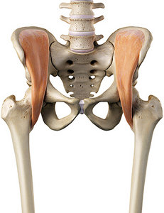 The iliacus has its origin on the iliac fossa and inserts on the lesser trochanter. Innervation is femoral L2-3. Action is to flex and laterally rotate the hip. It may be more prone to facilitation (neural upregulation) due to long periods of sitting. Physical therapist Shirley Sahrmann says if you have an inhibited muscle, look for a facilitated synergistic muscle in the pattern. A common pattern you find is inhibition of the psoas muscle and facilitation of the iliacus. However, the iliacus may compensate for other hip flexor muscles as well, depending on the pattern. Assess each muscle via palpation and muscle testing to determine strength:
- Tensor fascia latae
- Sartorius
- Rectus femoris
- Adductor longus
- Adductor brevis
- Adductor magnus
- Pectineus
- Gluteus minimus
- Gluteus medius (anterior fibers)
The iliacus is part of the deep front line (DFL) of fascia from Thomas Myers' Anatomy Trains. The DFL is a fundamental stabilization mechanism of the body. The iliacus has the same muscle-fiber orientation as the quadratus lumborum, a functional antagonistic muscle. If it connects to it, then it patterns to it.
A common relationship is an overactive iliacus compensating for weakness in the quadratus lumborum. Assessing movement can begin to reveal this possible relationship. An example is the overhead squat. Have the patient stand with feet shoulder-width apart with hands overhead in a "Y" pattern. Instruct them to go slow, squatting as deep and low as they can go. If they tuck tight into the hips, bringing the belly toward the thighs, it may indicate an overactive iliacus.
The quadratus lumborum muscles are extensors of the spine. If a person has inhibition in the QLs, they have difficulty maintaining extension in an overhead squat and compensate by taking the path of least resistance, tucking forward into flexion.
Palpation of a facilitated iliacus may illicit the "jump sign" response. Extreme tenderness is common with an overworked iliacus. Some patients may have a nervous system protective response and start laughing when you palpate a tender area. Since the iliacus sits deep, you need to passively flex the hip up beyond 90 degrees and passively adduct to reach it. Muscle knots in the iliacus can cause pain in the hip and groin region.
Muscle test the iliacus. It should lock and hold with testing. With the patient supine on the table, flex their hip beyond 90 degrees with slight abduction and external rotation. Instruct patient to bring knee toward the outside of the same-side rib cage. Resist motion on top of the thigh. Test one side, test the other side, then retest the first side. They should all be strong; no weakness.
Corrective Interventions
- Release the iliacus muscle through soft-tissue manipulation of choice.
- Adjust any subluxations discovered in joints through your palpation / manual testing.
- Teach the patient to actively stretch the iliopsoas complex. A lunge or the "warrior pose" movements in yoga are common ways to induce a stretch which works well, as long as the lumbars are not allowed to fall too far forward in the lunge and the pelvis is kept square to the front leg.
- Supine hip drill: Patient lies on their back with arms by their side, palms up. Keeping heel on the ground, slide heel toward the buttock (one leg at a time), flexing the knee. When foot is flat, lightly push the foot into ground for 4 seconds to contract the glute max complex. Next, instruct patient to lift their leg off the ground into a single-leg-stance position (hip flexion) and hold for 4 seconds; then return foot to the ground. Let the bent leg fall out into hip abduction. Control the eccentric phase down. When in maximal comfortable abduction, keeping the outside of the foot on the ground, slide foot back toward the starting position. Both legs should now be straight again. Repeat on the other side.
(Repeat four times total.)
Proximal stability precedes optimal distal mobility. Assess the central pillar powerhouse for efficient patterning, beginning with the iliopsoas complex. Remember, it's not all about the psoas.
Resources
- Cook G. Athletic Body in Balance. Champaign, IL: Human Kinetics, 2003.
- Gibbons S. Clinical Anatomy and Function of Psoas Major and Deep Sacral Gluteus Maximus. In: Vleeming A, et al (editors): Movement, Stability and Lumbopelvic Pain: Integration of Research and Therapy, 2nd Edition. Edinburgh: Churchill Livingstone of Elsevier, 2007.
- Myers TW. Anatomy Trains: Myofascial Meridians for Manual and Movement Therapists. Edinburgh: Churchill Livingstone, 2001.
- Osar E. Corrective Exercise Solutions to Common Hip and Shoulder Dysfunction. Chichester: Lotus Publishing, 2012.
- Sahrmann S. Diagnosis and Treatment of Movement Impairment Syndromes. St. Louis: Mosby, 2002.
- Weinstock D. NeuroKinetic Therapy: An Innovative Approach to Manual Muscle Testing. Berkeley, CA: North Atlantic, 2010.
- Yoshio M, Murakami G, Sato T. The function of the psoas major muscle: passive kinetics and morphological studies using donated cadavers. J Orthopedic Sci, 2002;7:199-207.
Click here for more information about Perry Nickelston, DC, FMS, SFMA.