


As chiropractors, we tend to focus on the spine, and rightly so. Many problems our patients face can be corrected by manipulating the correct spinal level. However, some pain can persist despite our best efforts, and I'd like to suggest a cause – and correction – for some of those problems.
Anatomy
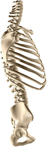
Twelve pairs of ribs articulate with each of the 12 thoracic vertebrae. Typically, the joint between the rib head and the vertebral body is shared between two vertebrae and their intervertebral disc. The rib also articulates with the transverse process of its vertebra, making three synovial joints for each rib. The rib is also actually attached to the intervertebral disc via a ligament. It ends in costal cartilage, with the top seven ribs attaching thereby to the sternum.
In addition to moving during breathing, the ribs are an attachment point for many muscles that are familiar problems in our patients, including the scalenes, erector spinae, quadratus lumborum in the back, and pectoralis minor in the chest.
 One of the most important joints related to the ribs is not a synovial joint at all, but the scapula sliding over the subscapularis as it retracts.
Signs of Joint Restriction
Restricted rib joints can manifest as localized pain, or the pain can follow the intercostal nerves around the thorax. Along with the painful deep breaths that sometimes accompany dysfunctional ribs, this condition can mimic a heart attack or pulmonary conditions. I have had patients go to the hospital first when faced with this pain.
With three joints per rib and all the muscular attachments, there is a lot that can go wrong with the ribs. Additionally, when the ribs do become restricted, their lack of movement can cause all those muscles to become irritated, resulting in pain seemingly unrelated to the cause.
The first rib is a special case due to its scalene attachments and their associated proximity to the brachial plexus. When you see radiating pain in the arm, check the first rib on the same side. A chronically restricted first rib (sometimes caused by poor sleeping posture) can cause scalene hypertonicity, which can radiate pain or tingling to the arm and hand.
One of the common problems seen with upper rib dysfunction is scapular protraction, or a forward shoulder posture. When the ribs are unable to move normally, the scapula cannot easily move over the rib cage posteriorly, and the shoulder becomes held in a forward position. Anytime posture is an issue, I start with posterior rib flexibility and find recent-onset problems can be solved quickly in this way.
Palpation
I use static palpation of the ribs to evaluate for restrictions. With the patient prone, contact the tubercle of the lower ribs T7-12, just laterally to the transverse process, and press P to A, evaluating joint mobility. Often a restricted rib will feel like it is protruding slightly, as if it is stuck in the posterior direction.
Upper ribs T2-6 can be felt more easily with the patient supine with their arms crossed. Hold onto the shoulder with your hand, rolling them toward you; and then allow the weight of their body to press the ribs into your curled fingers on the table.
The first rib can be palpated with the patient in a seated posture, pressing in at the angle of the neck, just anterior to the trapezius. Compare side to side to rule out normal from a restricted joint.
Adjustment
The lower ribs are adjusted in a prone position, contacting the ASIS with your hand and the base of your palm over the tubercle of the ribs. Pull up on the ASIS while keeping the ribs flat on the table.
The adjustment is less of a high-velocity thrust and more of a gentle application of weight through your arm. The line of drive should be as lateral as possible.
For ribs 2-6, a supine approach is best. Cross the patient's arms and lift the shoulder opposite your body, taking a contact over the costotransverse joint with your thenar eminence in an open hand. Release the shoulder and thrust your upper body through the patient's arms to compress the rib cage and move the posterior joints.
The first rib is adjusted in a prone position similar to a bench TM maneuver. Stand on the affected side and take a contact with the side of your 2nd MCP joint on the posterior 1st rib head.
Load your contact against the trapezius in a superior to inferior direction. Use your other hand to traction the patient's head toward you and rotate away, far enough to lock out the lower cervicals and upper thoracics. A high-velocity thrust in an inferior direction will release a restricted first rib joint.
In my clinical experience, properly adjusted ribs usually allow patients to breathe easier, reduce pain associated with the area, and most importantly, are an important part of proper posture.
Dr. Thomas French, a graduate of National University of Health Sciences, currently practices in Norwalk, Conn. Learn more at www.french-chiropractic.com.