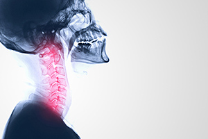
Would you like to know how to be able to diagnose the likelihood of traumatic cervical central disc herniation through X-ray (XR) alone? I have developed a practical technique for diagnosing traumatic cervical central disc herniation through a specific X-ray analysis. I can state almost without fail, this diagnostic method will show traumatic cervical central disc herniation that can later be confirmed by MRI. I have utilized this method of discovery through several hundred patient files.
The Follow-Up X-Ray: Clinical Value and Patient Buy-In
The follow-up XR study is a valuable tool in the diagnostic arsenal of the chiropractic physician treating traumatic motor vehicle accident (TMVA) patients. A follow-up (aka dynamic) X-ray is an X-ray taken within 3-4 weeks of the initial exam in patients with continued symptoms. The follow-up XR has the ability to demonstrate objective evidence of ligamentous sprain/subfailure and the likelihood of traumatic cervical central disc herniation when no evidence may have been observed on the original Davis series taken on the initial visit.1
 Indications for advanced imaging such as MRI can be supported by the results obtained through a follow-up XR study. In my experience in hundreds of cases, no follow-up XR has been denied for payment, and its use has been proven time and again to be successful in achieving maximum recovery for services rendered in treating the patient.
The follow-up XR study will help you uncover traumatic cervical central protrusion disc herniation injuries that may cause spinal cord injury (SCI) if due to severe sprain.2 The SCI may demonstrate varying degrees of sensory disturbance objectively and subjectively. Paresis and temperature impairment can occur and may be demonstrated upon the initial physical examination.2-3
These patients frequently have no initial extremity symptomatology or none extending through the inflammatory period of healing.4 Central disc herniation can contribute to spinal cord injury (SCI), anterior cord syndrome and central cervical cord syndrome.5-6 In a study by Flanders, et al., 54 percent of traumatic spinal cord injuries had associated disc herniations.7
Follow-up XR results can be reliably obtained when the cervical flexion and/or extension projections move at least 30 degrees in either plane.8 I have observed a consistent restriction in the extension ROM frequently when a posterior or posterolateral disc herniation is present and later confirmed on MRI.
Pro Note: A useful patient-education discussion point when describing the usefulness of the follow-up XR to the patient is to have them envision the popular building block game Jinga. In this application, envision the Jinga blocks wrapped tightly with plastic sheeting. This is what is happening when there is muscle spasm and guarding.
Describe to the patient that as the muscles heal and you unwrap the Jinga blocks, the block (i.e., spinal segment) is able to move without earlier restriction. Croft describes this as happening to the patient's spine as the muscle spasm and muscle guarding is diminished or is completely resolved by the third or fourth week post date of injury (DOI).9
Tips for Ensuring Accurate Measurement
Reliably measuring and understanding the clinical importance of the cervical neutral, flexion and extension images of the follow-up XR are critical. Noting the measurements in the radiographic report is also of significant importance.
For accurate measurements on plain film, I have been fond of the use of a rolling ruler that measures in millimeters (mm). Position the perpendicular black line against the very back edge of the vertebra below the vertebra you are comparing for the respective anterolisthesis or posteriorlisthesis. To determine the millimeter offset of the vertebra just above as the scale indicates may suggest a change in clinical management9 (to be discussed in the second part of this article).
With digital radiography, the process becomes a little more difficult. With digital XR, utilize a metallic marker of a known dimension to be placed on the bucky in the open space within the collimation. You then resize the picture to the pre-placed metal marker on the bucky after exposing the three images. When proper scaling has been achieved, listhesis measurements can be made accurately and then noted in the radiology report.10
Challenges and Other Considerations
There are a number of pitfalls in measuring the follow-up XR to review. Always measure the most posterior representations of the vertebra.9 It is extremely important to have good films. However, digital XR makes it possible to change contrast easily if the images are too dark or too light, in order to ensure you identify the true posterior aspect of the adjacent vertebra to the one you are measuring.
The measurement of angulation between two suspected hypermobile segments should be assessed. This measurement should not exceed 11 degrees of angulation. The technique for determining an instability in this manner is covered elsewhere.11
For an office that does not have on-site radiology, follow-up XR studies can be ordered at most imaging centers. Radiology studies contain a technical and professional component. You would be able to re-review the studies at your office and prepare a report noting the measurements as a medical decision- making (MDM) component that would satisfy that element of the correct level of E/M coding.12
References
- Herkowitz H, Rothman R. Subacute instability of the cervical spine. Spine, 1984;9(4):348-357.
- Rumboldt Z. Clinical Imaging of Spinal Trauma, A Case Based Approach. Cambridge, U.K.: Cambridge University Press, 2018: p. 73.
- Evans R. Neurology and Trauma. Oxford University Press, Inc., 2006: p. 311.
- Staller R. "Cervical Herniated Disc Symptoms and Treatment Options." Spine-health.com; last updated July 18, 2019. Click here to read
- White A, Punjabi M. Clinical Biomechanics of the Spine. Philadelphia: J. B. Lippincott company, 1978: p. 214.
- Rumboldt Z, Op cit.
- Evans R, Op cit, p. 266.
- Como J, et al. Cervical spine injuries following trauma. J Trauma, September 2009;67(3):651-9.
- Foreman S, Croft C. Whiplash Injuries: The Cervical Acceleration/Deceleration Syndrome, 2nd Edition. Philadelphia: Lippincott Williams & Wilkins, 2002: pp. 53-54.
- Ribeiro JP. "X-Ray Calibration: How to Scale Orthopedic Images for Templating." PeekMed Blog, Sept. 13, 2019. Click here to read
- White A, et al., Op cit: p. 225.
- "Q/A: Can I Bill a Review of X-Rays?" Chirocode Institute, Feb. 1, 2019. Click here to read
Dr. Troy Freiheit, a 1987 graduate of Northwestern Health Sciences University, operates private practices in Scottsdale and Phoenix, Ariz. He is also the founder of continuing-education provider PI Pro; for more information, contact Troy at
.


