


Iliac Flare Patterns - Inflare and Outflare
Most of us are familiar with iliac rotation in the transverse plane, which the Gonstead method calls IN or EX ilium. I use a different nomenclature, looking at the ilium from the front, calling this an external flare (IN in Gonstead) or an internal flare (EX in Gonstead). I evaluate and correct these conditions with the patient supine.
My evaluation is quite simple: Contact the ASIS bilaterally. On the side that feels restricted, push the ASIS into internal rotation, then pull the ASIS into external rotation. It will either move easily or feel restricted in one direction. Your other hand is used to stabilize the opposite side. Rely on the initial response testing (IRT) style of motion palpation (www.chiroweb.com/archives/19/16/12.html). You will feel stiffness or lack of give at the beginning of the motion. You will often find tenderness at the medial inferior border of the ASIS in a flare. I like using the ASIS for this evaluation, because it's at the end of a long lever, which makes it easier to use than the PSIS to test this motion. The standard muscle energy evaluation uses a visual assessment of position, looking at the ASIS position in relation to the midline of the body, observing whether the ASIS is medial or lateral. I find this difficult to do, as I am much more kinesthetic than visual.
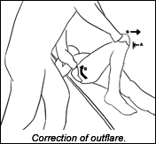
Correction of the outflare, in which the ASIS resists internal rotation, is simple. The patient is supine, with the bent leg standing. Flare corrections are long lever corrections, using the knee and femur to move the ilium (and thus the IS joint). The inferior hand is on the patient's knee; the superior hand is on the posterior lateral ilium, to monitor motion. Push the bent leg medially until the pelvis begins to move. At this point, have the patient push isometrically, laterally against your hand, for three to five seconds (arrow A in the image below and to the left). As the patient relaxes, take the knee further medially, into the receding barrier (arrows B). Repeat 2-3 times.
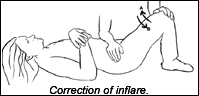
For an inflare, in which the ASIS resists external rotation, your correction is the opposite of the above process. Sit or stand on the opposite side of the restriction. You'll need to use the forearm or elbow of one arm to stabilize the near-side ASIS, while your palm monitors the involved ASIS. Your other hand brings the knee laterally until the involved side ASIS begins to move. Have the patient push medially (arrow A), isometric against your resistance for three to five seconds. As the patient relaxes, take the knee further laterally (arrow B), which, in turn, moves the ASIS laterally toward the new barrier. Repeat two to three times.
Ilio-Sacral Separation
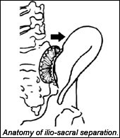
I've talked about how the SI can become hypermobile. This is another clinically significant subluxation pattern that really involves a combination of hypermobility and restriction. What are the biomechanics of this lesion? It involves a disruption of SI joint integrity. As with the iliac shears (upslip and downslip - refer to www.chiroweb.com/archives/21/03/08.html), SI hypermobility is a nonphysiological lesion, one to which the body cannot adapt. Chauffour, in his new text, Mechanical Link, talks about a functional diastasis, wherein two bones are separated and resist motion back toward proper connection. Chauffour states that these lesions "... are difficult to correct with classical techniques. The worst choice would be a thrust type of manipulation, which, contrary to its intention, often increases the instability of the diastasis." The concept of a functional diastasis is an extremely important, clinically significant one. It can be applied to the distal radio-ulnar joint; the distal tibiofibular articulation; the A-C joint; the pubic symphysis; and the IS joint.
Assess SI hypermobility with the patient prone. Your palpating hand has to start about two inches or more lateral to the PSIS to get enough tissue slack to assess the condition. Begin by sinking posterior-anterior into the gluteal tissues, then push medially toward the IS junction. Test at the S1, S2 and S3 levels. In an IS separation, the ilium, at the significant level, will resist your medial motion. The other finding here is exquisite tenderness. The confusing aspect is that this tenderness can exist in any SI pattern, not just IS separation.
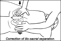
Here's the correction: Have the patient lie prone. Place your active hand, using either a thenar or hypothenar contact, on the tender, restricted spot on the ilium. The opposite hand takes a mirroring contact, on the contralateral ilium, also pushing medial. The whole of the pelvis is between your hands. Push medial with both hands, adding your three-dimensional components, finding the inferior-superior and clockwise-counterclockwise rotation directions that further load the barrier. Once you've found the exact maximal direction of resistance, remember to back off the barrier a bit, then listen and follow as the tissues release. You can also use recoil, either alone or along with the "engage, listen, follow" (ELF) correction. On large, bony areas such as this, I'll often use a percussive instrument, which helps get motion into these deep, rigid tissues. I tend to use this technique on the opposite side of the ilium, feeling the pulse come through to my main contact. The device seems to speed the release, and allows me to use less force.
Sagittal Rotation, Posterior-Inferior and Anterior-Superior Ilium
Posterior-inferior (PI) and anterior-superior (AS) ilium are also referred to as ilium vulgaris, the common iliac distortion. Typically, they are corrected over and over, far too often. From my experience and training with the osteopaths, this subluxation pattern is almost always a compensation, rather than a primary pattern. If you are correcting PI or AS ilia repeatedly, you are not getting to the root cause. You may even be contributing to hypermobility. When these show up, I always correct everything else I find around the pelvis and lower extremity, and then recheck the sagittal rotation. Usually, it will have spontaneously corrected. If sagittal rotation is a primary restriction, here's how to find and correct it.
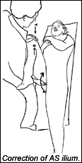
First, let's briefly review diagnosis. In a right AS ilium, the right ASIS will have moved inferior, and the right PSIS superior. Both the ASIS and the PSIS will resist motion in the corrective direction. If the lesion is significant, the SI will palpate as restricted on that side.
For an AS ilium, I use a supine "muscle energy" technique. My inferior hand is on the ischial tuberosity; my upper hand is on the bent knee. On the restricted side, I flex the bent leg until the pelvis starts to move. I then have the patient push toward me (extending his or her hip from full flexion) isometrically for three to five seconds (arrow A, above right). I'll resist the patient with my other hand, or with my shoulder, reminding him or her not to push too hard. As the patient relaxes, I can move the whole of the pelvis further into a posterior rotation (arrow B). I repeat this two to three times. In this instance, I use the hip to rotate the ilium posterior on the sacrum.
I rarely end up correcting a PI ilium. This seems to show up most frequently on the hypermobile side, or be a compensation of some kind. But if it truly is the key restriction, here's how to correct it: I use muscle energy, as it is gentle and effective. If necessary, I add a thrust. I'll have the patient side-lying, and set him or her up as if I'm going to do a diversified thrust for a left PI ilium. The patient is right-side-lying, with the left leg "hook-lying." I've got one hand on the ilium, and the other on the patient's bent knee. Once he or she is in the position that begins to open the joint, instead of thrusting, I'll have the patient push the leg up toward the ceiling, against the resistance of my knee. He or she pushes gently for three to five seconds, then relaxes. As the patient relaxes, I open the joint a bit further with my ilium contact. I'll repeat two to three times, then palpate the joint to see if it has freed up; if not, I may add a thrust, which is easier to do after the muscle energy methods have loosened the surrounding muscles. Again, it's worth noting that I rarely use this technique.
As many of the great chiropractic teachers have emphasized over and over, sacroiliac joint integrity is critical. Sacro-occipital technique has proven invaluable in furthering our understanding of the importance of low-force correction of this chronically unstable area. Mobility and stability of the SI region is critical for proper gait and overall neuromuscular health.
Next month, I'll complete the IS chapter with a look at the pubic symphysis and some fundamental rehab exercises for the sacroiliac region.
References
- Chouffour P. Mechanical Link, North Atlantic Press, 2002.
- Rex L. Introduction of muscle energy techniques (course notes), Edmonds, Wa., Ursa Foundation; 1996. p. 244-249.
- Greenman P. Principles of Manual Medicine, 2nd edition. Williams and Wilkins, 1996.
Click here for more information about Marc Heller, DC.