All of us who treat patients whose injuries led to them bringing a legal action against another party are likely to engage in debate as to the case's merits. Your duty as a treating doctor is to minister to the patient, providing optimal health care in order to achieve the best possible result.
Of course, all of this would be a lot easier if we didn't have to contend with defense lawyers. But, like taxes, they are a fact of life. As a result, your role is not only to cogently present the facts, but also to mute the myth, dispel the dogma, suspend the superstition, jettison the junk science, and basically disabuse the other forms of medical misadventure and legal legerdemain, including, I might add, accident reconstruction (although that is a subject unto itself).
The defense lawyer's job is to do whatever they can to reduce the defendant's exposure. They might try to deny liability from the start, essentially claiming the event was not their fault in the first place. When this option is not available, the next best strategy is to deny an injury occurred. A variant of this tactic is to argue that a plaintiff's current pain or impairment is the result of a pre-existing condition and not the event in question, be that a motor vehicle collision (MVC), an altercation or a fall.
This review will look at some popular defense tactics and methods of gulling juries, arbitrators and judges into accepting the defense theories based on myths and misconceptions related to imaging studies of the human spinal column.
Discounting Real Findings
This comes in two primary flavors. The first is to deny the significance of plain radiographic findings, a tactic that often starts with hospital studies. Hospital radiologists typically look at five-view studies (and sometimes the very low diagnostic quality cross-table laterals) to rule out serious pathology such as fracture and dislocation. Many also will note the absence of subluxations (the medical subluxation, of course) and imply there is no obvious ligamentous subfailure or plastic deformation. However, muscle spasm often will mask instability in the acute setting and definitive evaluation requires, at minimum, two additional bending views.1,2
Likewise with the cervical curve. The first logical argument is that without pre-injury films, you can't know whether the patient might not have had the same loss of curve before the injury. While that is undeniably true, studies by Gore revealed only 9 percent of the asymptomatic and atraumatic population have this abnormal configuration or anatomical variant, so the (91 percent) probability is, assuming the patient did not have a history of significant neck injury or prior neck pain, that the curve was normal before the injury.3,4
The second argument is that there is no relationship between the curve and the clinical state. Bohrer, et al., studied cervical spine flexion patterns in neck trauma.5 They felt that no flexion angle (i.e., straight spine) is consistent with more severe soft-tissue injury and that a single flexion angle or kyphosis probably indicates a grade I or II posterior ligamentous injury. Another group compared insidious onset neck pain and whiplash neck pain groups and found kyphotic configurations more common among the latter,6,7 suggesting a traumatic etiology.
The other primary flavor in terms of discounting real findings is to argue the significance of MRI findings of disc herniations. This also comes in two variants. In one, the opposing medical expert will argue that about 50 percent of asymptomatic people have a herniated disc, insinuating that the finding in the plaintiff may be nothing more than a red herring. In the other, the expert will argue that the radiologist overread the study and suggest the so-called herniation is really only a bulge. In this instance, I've seen some experts characterize even 5 mm herniations as "bulges."
The 50-Percent Solution: It is commonly believed, by a large number of doctors - and by nearly all lawyers and insurance people, it seems - that among the asymptomatic public at large, as many as 50 percent have at least one disc herniation. The genesis of this misconception can be traced back to a paper that appeared in the journal Spine in 1984.8 The authors used CT scanning to look at the lumbar spines of a number of adults who did not have back pain. Radiologists were told to read the studies and report any abnormalities they saw. In the over-40 age group, they found that about half showed some abnormality. However, many of these abnormalities were things other than disc herniations, including benign anomalies such as osteophytes.
Disc herniations were found in only 19 percent of the under-40 age group. Yet someone misinterpreted the results of this study as showing that 50 percent of all individuals (including those younger than 40) had at least one disc herniation in the lumbar region. And before long this misinterpretation was transmogrified into reality, popularized mostly by defense attorneys and their insurance company patrons eager to marginalize the findings of disc herniations in plaintiffs' backs. I can't even count the times I have been cross-examined by defense attorneys who brought this up. Naturally, I am always happy to disabuse that myth in detail, and I suspect those attorneys regret having broached the subject.
Subsequently, this confusion was cleared up by some of the same authors of the original study, although by then, this myth had attained a life of its own. Even today, this "50-percent herniation" myth is a prevalent one. Nevertheless, in other reports of asymptomatic disc bulge/herniation, reported figures range from 4-28 percent and herniations are more prevalent among the elderly.9 Interestingly, Boden, et al., reported that the relative proportions of "major abnormal findings" in the 20-39 age group and 40-59 age group were later shown to be very similar at about 20 percent; the 60-80 age group had markedly more abnormal findings at 57 percent, as one would expect.10 Thus, the rate of true positives (i.e., disc herniations that actually cause pain or other problems) is likely to be higher in younger people.
So, if we know that about 20 percent of the asymptomatic population has a lumbar disc with some degree of herniation, can this statistic safely be extrapolated to the cervical spine? Many lawyers and expert witnesses seem to believe so, even though any scientist worth their salt knows intuitively that it is dangerous to extrapolate beyond the available data. But the answer comes again by way of Boden, et al.,11 who found the incidence of herniation in the asymptomatic cervical spine to be only 8 percent. Only 2 percent had a bulging disc and 9 percent had foraminal stenosis or constriction at the point where the nerve root exits from the spine. Overall, the finding of "major abnormalities" in this group of subjects was only 19 percent. Yet degeneration was found in 37 percent of cervical spines (55 percent of the lumbar spines from their other study). And most recently, the incidence of cervical herniation among a group of 100 uninjured persons making up a non-whiplash control group was found to be only 4 percent.12 I have analyzed this cervical disc herniation literature elsewhere.13
An uncomfortable problem that is often ignored by clinicians and lawyers, but which is quite familiar to epidemiologists, is the "apples and oranges" mixing of populations. For example, if 19 percent of the asymptomatic population has one lumbar disc herniation, would we find the same proportion among a population of low back pain sufferers? If not, the meaning of the findings of a herniation in those two populations would be disparate. In other words, all of the disc herniations in the asymptomatic population could be thought of as false positives or type I errors. But in a symptomatic population, some of the herniations would undoubtedly represent true positives. Thus, there is no justification for extrapolating across populations. If your patient comes from a population of low back pain sufferers, the prevalence of disc herniation among non-back-pain sufferers is irrelevant.
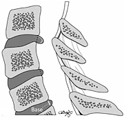
 This drawing illustrates three common lesions of the intervertebral disc: a bulge, a protrusion and an extrusion. The preferred term for the latter two is herniation.
So, having answered the question concerning the prevalence of disc herniation among the nontraumatic and asymptomatic population, we also might ask this question of the symptomatic population. What proportion of neck pain suffers have these lesions? In a recent study of neck pain sufferers, it was reported that only 17.3 percent of the C5-6 levels were deemed normal.14 This compared to 93.6 percent of the C2-3 spaces deemed normal in this series. "Protrusions/bulges" were found in 66.7 percent of cases and "extrusion" in 16 percent at the lower levels. But what precisely do these terms mean?
The proliferation of the disc terminology argot of radiologists eventually prompted the North American Spine Society (NASS) to establish a nomenclature development group or task force consisting of itself, the American Society of Spine Radiology (ASSR) and the American Society of Neuroradiology. The resulting document was endorsed by the task force, the Joint Section on Disorders of the Spine and Peripheral Nerves of the American Association of Neurological Surgeons (AANS) and the Congress of Neurological Surgeons (CNS), as well as by the CPT (Current Procedural Terminology) and ICD (International Classifications of Disease) Coding Committee of the American Academy of Orthopaedic Surgeons (AAOS). This will serve as the new definitive terminology. If Dr. Evil wants to call a 5 mm disc herniation a "bulge," he is swimming against a pretty swift current now.
Bulging of the disc connotes less than 3 mm of extension beyond the normal margin. There have been many terms used in the description of herniation. We have herniated nucleus pulposus (HNP), but it is discouraged now. Ruptured disc is also discouraged, as was disc prolapse. Then there is the term disc protrusion, which represents a herniation in which the distance of protrusion is less than the width of the base. And finally we have disc extrusion, in which the distance of protrusion exceeds the width of the base. In the final analysis, the terms of choice are bulge and herniation. And now you probably know more about this mysterious lexicon than most other physicians. Figure 1 illustrates the accepted new terminology.
Misattributing Pain to "Normal" Pathology
Another common medicolegal misconception is that the presence of degeneration is generally always abnormal, likely to be painful and the likely source of a patient's pain (as opposed to the claimed injury).
The poor correlation between clinical states and degenerative spines is familiar to all doctors. Research confirms this.15 There may be some correlation in younger people, especially when there is loss of disc height and focal changes indicating trauma, but after the age of 40 years, correlation becomes less certain.16 But such a correlation is a relatively simple canard to foist on an unsuspecting jury. After all, words such as disease and degeneration imply pain to most people. But spondylosis deformans is a product of aging and will be found in about 50 percent of normal 30-year-old spines and in about 80 percent of 50-year-old spines. By age 70, approximately 70 percent of us have degenerative spinal changes in the cervical spine.17 Importantly, only about 20 percent of adults have chronic neck pain,18 making the lack of concordance between skeletal radiology findings and the clinical state rather plain. Intervertebral osteochondrosis (disc disease) is nearly as common, with signal changes from desiccation typically beginning in teenagers. It has a little more correlative power than spondylosis, with loss of disc height somewhat more often seen (OR: 2.1) among back pain sufferers.15 Matsumoto, et al., found disc degeneration to be their the most common observation of asymptomatic subjects on MRI, being present in 17 percent of discs of men and 12 percent of those of women in their 20s, and 86 percent and 89 percent of discs of men and women, respectively, over 60 years of age.19
Ruling Out Pathology With Imaging Studies
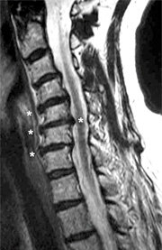
 Three-level anterior herniation with one-level posterior herniation (asterisks) in a female whiplash victim who now has constant moderate neck and shoulder pain.
The final medical misadventure is the peremptory dismissal of all potential soft-tissue or bony lesions based on radiographic or advanced imaging studies, usually MRI. Early animal experiments demonstrated many of the spinal soft-tissue lesions produced during whiplash trauma were invisible to plain film.20,21 Studies in humans have demonstrated that rather significant findings at autopsy are not present on plain films and, in some cases, MRI.22,23 The latest among this group of studies describing occult injuries in motor vehicle trauma is from Stabler, et al.24 The authors reported: "Lesions of the facet joints were detected only indirectly on the basis of depiction of fluid in the joint. Direct depiction of the ruptured apophyseal joint capsule was almost impossible." This is a particularly significant finding coming from the likes of fellow San Diegan Don Resnick, the author of the mammoth five-volume tome, Diagnosis of Bone and Joint Disorders, regarded by many to be the preeminent authority of bone and joint radiology today.
When Resnick can't find something on an MRI or radiograph, it generally isn't there. Like previous authors, these authors found that only a minority of serious injuries in the spine were fractures. Three of the lesions they found (11 percent of the total of 28 lesions) were fractures. Of special note, two of them were not visible using plain radiographs. Radiographs, in fact, depicted only one of the 28 lesions found. And only 11 of the 28 lesions were initially found on MRI. Seventeen were found only after correlation with the pathologic examination of the specially prepared 3-micrometer slices of these spinal segments. Bottom line: The resolving power of X-ray and MRI is insufficient to visualize the full spectrum of trauma. And that's if you always know what to look for.
Discography studies have demonstrated that even morphologically intact discs can produce pain, so herniations or obvious structural changes don't give us the whole story. Jinkins, et al., demonstrated that anterior disc herniations also can be a source of pain.25 Based on the finding that normal cervical discs are innervated, I believe anterior herniations are also relevant in the cervical spine (Figure 2). In my experience, most radiologists don't even comment on these findings.
References
- Griffiths HJ, Olson PN, Everson LI, Winemiller M. Hyperextension strain or "whiplash" injuries to the cervical spine. Skeletal Radiol, 1995 May;24(4):263-6.
- Griffiths HJ, Kidwai AS, Wright WC. Hyperextension strain or whiplash injuries to the cervical spine „Ÿ revisited. J Whiplash Related Disorders, 2004;3(1):25-45.
- Gore DR. Roentgenographic findings in the cervical spine in asymptomatic persons. Spine. 2001;26(22):2463-6.
- Gore DR, Sepic SB, Gardner GM. Roentgenographic findings of the cervical spine in asymptomatic people. Spine, 1986;11(6):521-4.
- Bohrer SP, Cherr YM, Sayers DG. Cervical spine flexion patterns. Skeletal Radiol, 1990;19:521-5.
- Kristjansson E, Jonsson H, Jr. Is the sagittal configuration of the cervical spine changed in women with chronic whiplash syndrome? A comparative computer-assisted radiographic assessment. J Manipulative Physiol Ther, 2002 Nov-Dec;25(9):550-5.
- Kristjansson E, Leivseth G, Brinckmann P, Frobin W. Increased sagittal plane segmental motion in the lower cervical spine in women with chronic whiplash-associated disorders, grades I-II: a case-control study using a new measurement protocol. Spine, 2003 Oct. 1;28(19):2215-21.
- Weisel S. A study of computer assisted tomography: I. the incidence of positive CAT scans in an asymptomatic group of patients. Spine, 1984;9(6):549-51.
- Kent DL, Haynor DR, Larson EB, Deyo RA. Diagnosis of lumbar spinal stenosis in adults„Ÿa meta-analysis of the accuracy of CT, MR and myelography „Ÿ review. Am J Roentgenol Radium Ther Nucl Med, 1992;158(5):1135-44.
- Boden SD, Davis DO, Dina TS, Patronas NJ, Wiesel SW. Abnormal magnetic-resonance scans of the lumbar spine in asymptomatic subjects. J Bone Joint Surgery, 1990;72-A(3):403-8.
- Boden SD, McCown PR, Davis DO, Dina TS, Mark AS, Wiesel S. Abnormal magnetic-resonance scans of the cervical spine in asymptomatic subjects. J Bone Joint Surgery, 1990;72-A(8):1178-84.
- Giuliano V, Giuliano C, Pinto F, Scaglione M. The use of flexion and extension MR in the evaluation of cervical spine trauma: initial experience in 100 trauma patients compared with 100 normal subjects. Emerg Radiol, 2002 9(5):249-53.
- D'Antoni A, Croft AC. Prevalence of herniated intervertebral discs of the cervical spine in asymptomatic subjects using MRI scans: a qualitative systematic review. Journal of Whiplash and Related Disorders, 2005;5(1):5-13.
- Arana E, Marti-Bonmati L, Molla E, Costa S. Upper thoracic-spine disc degeneration in patients with cervical pain. Skeletal Radiol, 2004 33(1):29-33.
- Pye SR, Reid DM, Smith R, Adams JE, Nelson KE, Silman AJ, O'Neill TW. Radiographic features of lumbar disc degeneration and self-reported back pain. J Rheumatol, 2004;31(4):753-8.
- McRae DL. The significance of abnormalities of the cervical spine. Am J Roentgenol Radium Ther Nucl Med, 1960;84:3.
- Fenlin J. Pathology of degenerative disease of the cervical spine. Symposium on disease of the intervertebral disc. Orth Clin North Am, 1971;2:371-87.
- Guez M, Hildingsson C, Nilsson M, Toolanen G. The prevalence of neck pain: a population-based study from northern Sweden. Acta Orthop Scand, 2002 Aug;73(4):455-9.
- Matsumoto M, Fujimura Y, Suzuki N, Nishi Y, Nakamura M, Yabe Y, Shiga H. MRI of cervical intervertebral discs in asymptomatic subjects. J Bone Joint Surg Br., 1998;80(1):19-24.
- Macnab I. Acceleration injuries of the cervical spine. J Bone Joint Surg Am., 1964 Dec;46:1797-9.
- Wickstrom J, Martinez J, Rodriguez R. Cervical pain. New York: Pergamon Press; 1972.
- Taylor JR, Taylor MM. Cervical spinal injuries: an autopsy study of 109 blunt injuries. Musculoskeletal Pain, 1996;4(4):61-79.
- Uhrenholt L. "Morphology and Pathoanatomy of the Cervical Spine Facet Joints in Road Traffic Crash Fatalities With Emphasis on whiplash -- A Pathoanatomical and Diagnostic Imaging Study." (PhD thesis) Aarthus, Denmark: University of Aarthus, 2007. 107 p.
- Stabler A, Eck J, Penning R, Milz SP, Bartl R, Resnick D, Reiser M. Cervical spine: postmortem assessment of accident injuries -- comparison of radiographic, MR imaging, anatomic, and pathologic findings. Radiology, 2001;221(2):340-6.
- Jinkins JR, Whittemore AR, Bradley WG. The anatomic basis of vertebrogenic pain and the autonomic syndrome associated with lumbar disk extrusion. Am J Neuroradiol, 1989;10:219-31.
Click here for previous articles by Arthur Croft, DC, MS, MPH, FACO.


