One morning in 1955, before air conditioning was in wide use, a Texas DC walked into his office and found it hazy with smoke. He quickly checked the most likely sources, such as the hydrocollator heaters and the electric wiring.
A similar confusion exists for the myofascial therapist. The confusion concerns the role of the spine in some patients' pain. We are taught a useful diagnostic principle: A patient's pain pattern can lead us to the muscle that's referring the pain (as every muscle refers pain in a pattern characteristic for that muscle).1,2,3 The relationships are highly reliable -- a patient's pattern of pain leads to the referring muscle as dependably as smoke leads to the fire from which it came.
Some patients' trigger points (TP), however, refer in unusual or eccentric patterns. Dr. David Simons reported palpating patients who exhibited unusual referral patterns.4 One of the patients he examined complained of knee pain and another complained of shoulder pain. To paraphrase Simons, "It didn't matter what TP I pushed on, instead of the pain going where I expected it to -- where it would in the typical myofascial patient -- the pain was always referred to the site of the patient's pain complaint: one patient's knee, the other's shoulder. It looked to me like there was a pain modulation syndrome where the referral was being redirected to a particular site."
When I first encountered these strange pain patterns in patients, I thought that treating the TP locally would relieve the pain, regardless of why it was distributed in an unusual way. In most cases, as I desensitized the TP, the patient's pain subsided -- but only temporarily. When the pain returned, however, the TP I had neutralized wasn't necessarily the source. Like Simons, I noticed that TPs in various other muscles would also refer to the same location.
For a period of months, I puzzled over this phenomenon. Finally, I realized the basis of the odd pain patterns. But I was embarrassed that as a DC, I had overlooked the obvious for so long.
In most every case, the eccentric pain pattern was in the dermatome of a lesioned spinal segment. The lesion might be some spinal pathology, a recurrent vertebral subluxation, or a stressed spinal area such as the thoracolumbar junction. Regardless of the location of the TP, it referred pain in some part of the skin innervated by that lesioned spinal segment.
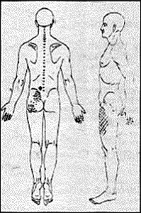
 Figure 1: The x: TP. Stippled area: classic referral pattern. Cross-hatched area: eccentric referral pattern (segmentally related to lesioned spinal segments).
Recently, doctors attending my Los Angeles seminar witnessed, firsthand, a clear example of eccentric myofascial pain. I was demonstrating how to palpate the lumbar paraspinal muscles with the elbow. I pressed my elbow deeply into a volunteer's lower back muscles and found a TP. She said the pain was referred to the front of her thigh on the same side. The classic pain pattern for the muscle I was compressing is the sacro-iliac (SI) and gluteal areas. Because of this, I assumed that her thigh pain was an eccentric distribution (in the dermatome of a lesioned spinal segment). The anterior thigh contains the dermatomes for the upper lumbar spinal segments, so I palpated them for fixations. I applied pressure to the spinous processes at those levels and she said they were sore. I then pressed the tip of my elbow into the multifidus muscles just lateral to the spinous processes, and these too were tender on both sides at L1, L2, and L3. I also pressed down over these segments and found that as a unit they where hypomobile. The pattern suggested that the involved vertebrae were subluxated. Figure 1 shows the location of the TP, its classic referral pattern, and the dermatomal pattern where her pain was referred.
Simons is right about this phenomenon involving a pain modulation syndrome. A TP is irritable enough to refer pain, but the pain isn't felt at its classic anatomical site. Instead, it's redirected by a lesioned spinal segment -- just as the DC's fan redirected the restaurant's smoke into his office.
The mechanism of eccentric patterns is called segmental facilitation and has been identified and clarified by Irvin Korr and other researchers. Their studies showed that lesioned spinal cord segments were "chronically hyperirritable and, therefore, hyperresponsive to signals reaching them from any source (emphasis mine) in the body."5 Koor6,7 injected salt solution into myofascial tissues to demonstrate that such irritants cause reactions in the dermatomes of lesioned cord segments. Myofascial TPs are probably the most common irritants that provoke reactions in lesioned segments. A lesioned segment acts as a "receiving site"8 for noxious signals from TPs. This site then broadcasts these signals as muscle contractions, sweat secretion, vasoconstriction, and pain
When your patient describes the pattern of his pain, palpate the muscle that normally refers that pattern. If you find a TP, treat it. Whether or not you find one, note the skin area the pain covers, and examine the spinal segment that innervates that dermatome. Check for subluxations, pathology, and postural stress -- the neuropathological component of these conditions may be a "fan" diverting the patient's pain. If the segment is lesioned, treat both the spine and myofascia for optimal therapeutic results.9
Refernces:
- Travell, J.G. and Simons, D.G. Myofascial Pain and Dysfunction: The Trigger Point Manual 1983.
- Lowe, J.C. The Purpose and Practice of Myofascial Therapy (audio cassette album) Houston, McDowell Publishing Co. 1989; Tape 3.
- Smolders, J. Trigger Point Charts Fjes 1984.
- Bennett, R.; Goldenberg, D.L.; Simons, D.G.; and Travell, J.G. "Panel on definitions and diagnostic criteria for muscular pain syndromes." First International Symposium on Myofascial Pain and Fibromyalgia, May 8, 1989.
- Korr, I.M. "Somatic Dysfunction, Osteopathic Manipulative Treatment, and the Nervous System: a few facts, some theories, many questions." Journal of the American Osteopathic Association February 1986; 86(2): p 110/98.
- Korr, I.M.; Wright, H.M.; and Thomas, P.E. "Effects of Experimental Myofascial Insults on Cutaneous Patterns of Sympathetic Activity in Man." Act Neuroveg. 1982; 23: p 329-355.
- Korr, I.M. "Experimental Alterations in Segmental Sympathetic (Sweat Gland) Activity through Myofascial and Postural Disturbances." Federation Proceedings 1949; 8: p 88.
- Korr, I.M. Lecture on Segmental Facilitation, Toronto, April 12, 1984.
- Lowe, J.C. "Treatment-resistant Myofascial Pain Syndromes." Soft Tissue Examination and Treatment by Manual Methods, Warren Hammer (ed.), Baltimore, Aspen Publishing Co 1990; in press.
Click here for previous articles by John Lowe, MA, DC.


