Dural tension is intimately involved with disc pain. When a disc bulges posteriorly, it can compress the dura directly. The dura, another mesodermally derived fascial structure, responds and adapts to all of the biomechanical changes in the surrounding tissues. One of the themes of the manual approach is that all fascial structures and mesodermally derived tissue can become restricted and be released by various forms of adjustments and/or soft tissue release work.
The dura is the external lining of the spinal canal; it is continuous with the meninges of the brain (think "cranial restrictions" and "cranio-sacral manipulation"). The dura is contiguous with the exiting spinal nerves, and the nerve sheath that protects the peripheral nerves is another fascial lining that begins at the dura.
Assessing Dural Tension
"Hamstring" tightness is not just a hypertonic muscle. If you perform a straight leg raise (SLR), you are assessing the tension in the sciatic nerve. In a classic disc herniation, you'll get quite a diminished SLR at 20-50 degrees of leg raise, causing shooting leg pain. In an internally disrupted disc, or any chronic lumbosacral problem with accompanying dural irritation, the SLR will also be diminished, but the result will be subtler. Instead of a clearly positive SLR, itwill be 10-30 degrees less than the opposite side, and you will elicit tightness in the hamstrings. This will often correlate with referred pain into the buttock and posterior thigh. When conducting the SLR test, remember the "soft barrier" concept. Lift the straight leg until you feel the beginning (minimum) resistance, not maximum. The beginning of the resistance is where you have engaged the tension in the nervous system, from the lumbar dura down into the sciatic nerve.4
Assessing excessive dural tension is often less clear than spinal subluxation. When should you suspect that the dura is involved? The dural attachments will be stiffer when the dura is irritated and pulling, so the sacrum and coccyx may be tender and restricted. The sacrotuberous ligament is fascially continuous with the sacrum and parallels the sciatic nerve; it will usually be tight and tender on the side of dural tension. The midline tenderness test3 will often be positive at two or more lumbar levels.
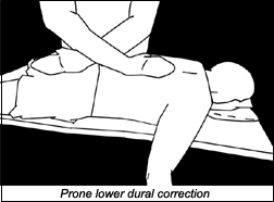
Here's a direct test of dural tension:5 The patient is lying prone on a bolster to eliminate the lumbar lordosis. The first step is to sink, rather than push, posterior to anterior, through the bony spine down to the dural level. You want to assess the deep structures, not the superficial fascia. This is a rather subtle skill, but it can be learned easily. Your lower contact is on the sacrum, taking it further into counternutation, and taking the sacral apex inferior and anterior. CAP
Your second contact is on the midthoracic spine, at the apex of the patient's kyphosis. Take the contact with crossed hands, sink, and then gently push apart. Your hands should not glide across the skin; they should feel for a give in the deep structures.
Correcting Dural Tension
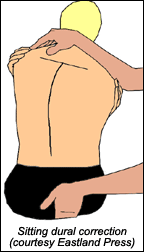
The correction follows directly from the assessment described above, tractioning the dura with your hands, with the addition of the "engage, listen, follow" (ELF) concept of three-dimensional simultaneous assessment and release. Another way of correcting the dura takes advantage of the fact that the dura becomes slack on spinal extension and tighter on flexion. With the patient sitting, contact the sacrum, coccyx, or both with one hand. Begin by lifting the coccyx-sacrum superior, engaging the dura, then "back off" slightly. Note that lifting superior is an indirect way of engaging; slacking, rather than stretching. If I am on the sacrum, I take a midline contact.
On the coccyx, contact the side where the tension is greater, just under the sacrotuberous ligament. Have the patient slump forward slightly, emphasizing the reversal, or rounding, of the lumbar lordosis. You will feel the tension develop in the spine; don't let the patient bend too far forward. Have the patient cross the arms, and side-bend him or her one way, then the other. Find the side-bending direction that increases the overall tension. Move the patient in this direction to the soft barrier, and feel the dura begin the stretch and release, over 20-90 seconds. Beginners will typically move the patient too far, both in flexion and lateral bending, going far beyond the beginning of the barrier. The sitting technique is especially effective when there are unilateral muscular bands, which often reflect unilateral dural tension. I recommend Barral and Crobier's text, Trauma, An Osteopathic Approach, both for the techniques and for a deeper understanding of how trauma impacts the dura. I have addressed the cervical dura, and introduced this conceptual model in a previous article.6
Using a vertebral distraction pump or manual axial distraction can also mobilize the dura. Add the dural correction by mobilizing the sacrum in harmony with the craniosacral rhythm, as described in my previous article.6
Release of the Sciatic Nerve Via Nerve Gliding
As chiropractors, we tend to work on the joints, and assume that we are releasing the nerve. Think "fascial linings!" We can also work directly on the fascial tension along the nerve itself. This notion is based on David Butler's nerve tension work. Mark Bookhout took these nerve tension concepts and integrated dural tension (cranial-based) concepts.7,8 Jean-Pierre Barral has recently developed similar techniques to correct peripheral nerve tensions.9
You can use a combination of nerve gliding with an externally applied fulcrum at the significant segment (such as a small ball, for self-mobilization), to free dural restrictions while the patient is gliding the sciatic nerve, by straightening the bent leg. This is particularly useful for sciatica or any dural tension affecting the sciatic nerve. I recommend that you do this as an in-office procedure first. This allows you to test whether it is effective for the patient, as shown by an immediate increase in his or her straight leg raise, and is a way to effectively teach the exercise. I teach my patients to use nerve gliding as a home exercise.
I am deliberately using the word "gliding," rather than "stretching." Overstretching can injure the delicate nerve tissue. If overdone, this exercise can further irritate or injure the sciatic nerve. Instruct the patient to be gentle in the raising of the leg; go only to the beginning of the nerve stretch/pain, then come back out.
In-office version: Find the most restricted and tender segment, as tested via lateral-to-medial pressure on the spinous process. The tenderness/restriction can be on the side of symptoms, or on the opposite side. With the patient supine, reach under the patient's body and across the midline. Your palm cups the middle of the spine and your fingers pull on the spinous process, from the opposite side toward the midline. Having found the most tender and restricted spinous, apply gentle pressure with your fingers, lateral to medial. Now, ask the patient to lift his or her thigh with the bent leg, and have him or her grasp with both hands behind the bent knee. From this position, the patient slowly and actively lifts the lower leg until tension is felt behind the knee or in the thigh. Next, lower the patient's leg. You are holding the spinous with an active isometric pressure. You'll feel the spine move into your fingers. Repeat 3-10 times. Recheck the patient's SLR for increased ease of lifting. When this mobilization works, it will increase the ease and range of motion in the SLR.
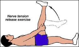
At-home exercise (described for the patient): As pictured above left, lie on your back, and grasp behind the knee. Support your head with a pillow if this makes you more comfortable.
Start by lifting your bent thigh, grasp behind the knee, and slowly straighten the lower leg. Let your leg down as soon as you start to feel a stretch. Keep your toes pulled back toward your shin; don't point your toes upward. As you lift your leg, you should feel tension in the back of your thigh or behind your knee. You should not feel sharp shooting pains in the leg or back. You can increase the tension by bringing the thigh slightly across toward the opposite thigh and holding it there with your arms, while you straighten your leg. Repeat slowly 10 to 20 times, 1-3 times per day. (Variation: You can do this with a small soft ball, such as a racquetball or hackeysack, behind your low back on one side or the other.) Be gentle; do not overdo this exercise, as it can be harmful if done too vigorously!
Addressing the dura and the sciatic nerve directly will further help your patients with chronic lower back issues and potential discogenic pain. Please remember to be gentle with these delicate nervous system sheath structures: Less force is always more effective when working with the dura and nerve sheath.
References
- Heller M. Correction of discogenic pain and midline tenderness. Dynamic Chiropractic, Oct. 6, 2003. www.chiroweb.com/archives/21/21/09.html.
- Heller M. Discogenic pain: diagnosis and treatment. Dynamic Chiropractic, Sept. 1, 2003. www.chiroweb.com/archives/21/18/08.html.
- Discogenic pain: inflammation and exercise. Dynamic Chiropractic, Nov. 17, 2003. www.chiroweb.com/archives/21/24/07.html.
- Butler D. Mobilization of the Nervous System, 1995, Churchill Livingston.
- Barral JP, Crobier A. Trauma, An Osteopathic Approach, Eastland Press, 2001.
- The dura mater and the spinal cord. Dynamic Chiropractic, Jan. 14, 2002. www.chiroweb.com/archives/20/02/06.html.
- Bookhout M, Greenman P. Principles of Exercise Prescription, Butterworth-Heinemann, 2003.
- Bookhout M. Exercise as an Adjunct to Manual Therapy, 1998 (seminar).
- Visceral Approach to Trauma and Whiplash, level 1 and 2, seminars, 2002, 2003.
Click here for more information about Marc Heller, DC.


