How Does Low Back Injury Occur?
Injury occurs when external load exceeds the failure tolerance or strength of the tissue. In particular, low back injury has been shown to result from repetitive motion at end-range. According to McGill, it is usually a result of "a history of excessive loading which gradually but progressively reduces the tissue failure tolerance" (McGill 1998).
Disc injury has been shown to be related to three factors: first, full end-range flexion in younger spines (due to higher water content) (Adams, Hutton 1982; Adams, Hutton 1985; Adams, Muir 1976); second: repetitive end-range flexion loading motion in excess of 20-30,000 cycles (King 1993; Gordon et al., 1991); and third, epidemiological association between disc herniation and sedentary sitting occupations (Videman et al., 1990).
Certain times of day are the most vulnerable for the back. Disc bending stresses are increased by 300% and ligaments by 80% in the early morning (Adams MA et al., 1987). McGill has reported that after just three minutes of full flexion, subjects lost half of their stiffness (McGill, 1989).
How Does the Body Resist Injury?
Support and stability to the low back arise from muscles. Surprisingly, the one muscle that is highly active during flexion, extension and lateral bending tasks is the quadratus lumborum (McGill, 1996). Its architecture is ideally suited to be a stabilizer since it attaches each transverse process to the more right pelvis and rib cage, facilitating a bilateral buttressing effect for the vertebrae (McGill, 2000).
Interestingly, it is not strength but coordination between agonist and synergist muscles which plays a pivotal role in resisting injury. Sparto et al., report that spinal loading forces increased during a fatiguing isometric trunk extension effort without a loss of torque output (Sparto, 1997). Torque output remained constant because as the erector spinae fatigued, substitution by secondary extensors such as the internal oblique and latissimus dorsi muscles occurred.
Agonist-antagonist muscle coactiva-tion is an important mechanism in preventing spine injury. Cholewicki et al., examined the theory that antagonistic trunk muscle coactivation is necessary to provide mechanical stability to the lumbar spine around a neutral posture (Cholewicki, 1997). The authors found that antagonistic muscle coactivation increased in response to increased axial load on the spine.
Correlation of Motor Control Dysfunction with Low Back Problems
Altered activation sequences and ratios between synergist muscles has been correlated with lower back pain (LBP). A decreased activation of the transverse abdominus/oblique abdo-minals relative to the rectus abdominus was correlated with LBP (O'Sullivan P et al., 1997). Control subjects were able to preferentially activate the internal oblique and transverse abdominus without significant rectus abdominus activation, whereas LBP patients could not do this.
Hodges and Richardson showed that delayed activation of transverse abdominus during arm movements distinguishes LBP patients from normals (Hodges, Richardson 1996).
Decreased endurance of the trunk extensors has not only been shown to correlate with pain, but to predict recurrences and first time onset in healthy individuals (Biering-Sorensen F 1984; Luoto et al. 1995). The multifidus in the low back has been shown to be atrophied in patients with acute low back pain (Hides et al., 1993). Recovery from acute pain did not automatically result in restoration of the normal girth of the muscle (Hides et al., 1996). However, spinal stabilization exercises successfully did rebuild the muscle's size and resulted in reduced recurrence rates in the future.
Injury Prevention Advice
Tissue conditioning must occur as exposure to load increases. If rest time is inadequate, training is absent, or forces are excessive, then injury will eventually result. Too little or infrequent exposure to external load and conditioning never raises the tissue failure tolerance. In other words too much, frequent or prolonged exposure, and adaptation can't keep pace.
Knowledge of when an injury is most likely to occur can also influence what we do. Early morning or after prolonged sitting are particularly vulnerable times. Reilly et al. (Reilly et al., 1984) showed that 54% of the loss of disc height (water content) occurs in the first 30 minutes after arising. Disc bending stresses are increased significantly in the morning (Adams, et al. 1987). After even a brief period of sitting or stooping, end-range protective joint stiffness is compromised (McGill, 1999). Even after 30 minutes of rest, residual joint laxity persisted! Therefore, avoidance of high-risk activities early in the morning or after sitting or stooping in full flexion is crucial to injury or reinjury prevention.
Suggestions to teach workers to lift with their knees, not their backs, are overly simplistic. Most workers have learned various techniques to avoid injury which are inconsistent with this advice. Better advice is consistent with the following principles: Avoid end-range motion. Rotate jobs to vary loads. Allow frequent rest breaks. Keep loads close to the spine (McGill, Norman, 1993).
Rehabilitative Exercises
Axler and McGill have demonstrated that muscle output and spinal load can be measured for a variety of exercises (Axler, McGill, 1997). Muscle output is determined as a percentage of maximum voluntary contraction ability (MVC) and spinal load as a measure of spinal compression and shear forces. Ideal exercises have a high ratio of muscle challenge to spinal load. Such analysis gives surprising data about common exercises which are prescribed for low back pain. For instance, spinal load is not different during situps with knees bent or straight. In either case, the load is over 3000N and therefore should not be prescribed in the low back recovering population (Axler, McGill 1997; McGill, 1995).
Safe back principles have emerged from rigorous analysis of biomechanical and kinesiological aspects of spinal function (McGill, 1998b; Liebenson, 1999). By emphasizing endurance training of key spinal stabilizers, positive outcomes have been achieved (Hides et al., 1996; Timm et al., 1994; O'Sullivan et al., 1997b). These principles can be summarized into a user-friendly approach which will enhance clinical application and patient compliance (See Table I).
Table I: Safe Back Exercise Menu
- limbering
- kinaesthetic awareness of "neutral posture"
- the big three:
• anterior
• lateral
• posterior - flexibility for peripheral joints:
• ankle
• knee
• hip - cardiovascular fitness
Patients begin with a simple warmup involving the cat/camel exercise on all fours. This should be performed first thing in the morning and then prior to endurance training exercises. Between 8-10 repetitions are all that are needed. Think of the cat/camel as a limbering, rather than stretching, movement.
After performing the cat/camel, the patient is ready for simple exercises to improve the endurance of the abdominal and back muscles. Incorporated into each exercises should be a motor control component teaching "neutral spine" kinaesthetic awareness. The main emphasis of rehabilitative exercise is to train "inner range" endurance. This requires kinaesthetic awareness of "neutral spine" postures and regular sustained training of this motor control against a variety of challenges. Multiple reps of 5-6 second holds should be performed on a daily basis. According to Manniche et al., up to three months may be required to achieve a long-lasting beneficial effect (Manniche et al., 1991).
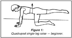
The back muscles can be tested in the modified Sorensen position. Normative data has been established (Alaranta, 1994). They can be trained with the quadruped single leg raise (see Figure 1). Always ask the patient to draw their navel in toward their back to coactivate the deep spinal stabilizers. This can progress to quadruped opposite arm and leg raises and the superman exercise on the gym ball.
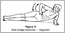
The side muscles (quadratus lumborum and internal oblique abdominals) can be trained in the side bridge position. They can be tested utilizing a side bridge. Normative data has been established (McGill et al., 1999b). The normal ratio of static back extensor endurance (Sorensen test) to side bridge endurance is .73.
Training starts with the side bridge supported on forearm, knees and ankles (see Figure 2). The next progression is with legs straight using only the lower arm and ankles for support. A final progression is to perform the horizontal bridge on ankles and, without lowering the body, roll on to the other forearm. This adds a motor control or balance element to the exercise. Remember: each side bridge exercise can be intensified by tightening the abdominals by drawing the navel in toward the back.
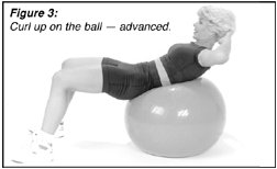
The abdominals can be tested for endurance in a variety of ways (Alaranta, 1994; McIntosh et al., 1998; McGill, Liebenson, 1999). They can be trained with trunk curl-ups or "dead bug" exercises. Progressions can include adding twists and/or using the gym ball (see Figure 3). The gym ball, because of its labile nature, results in greater activation of the transverse abdominus and oblique abdominal muscles.
Always remember to instruct your patient if they experience discomfort. The cat/camel exercise is their "first aid" exercise. A little bit of discomfort with the other exercises is all right, as hurt does not necessarily equal harm. The road to recovery is through activity. Patients are educated that if their back gives them trouble, it is a sign that it is not strong or supple enough to do the work required. The cat/camel and other exercises shown here are safe for the back and will help recondition the "weak link."
Acknowledgements
I would like to thank professor Stuart McGill for his many contributions, upon which much of this article has been based.
References
Adams MA, Hutton WC. Prolapsed intervertebral disc: a hyperflexion injury. Spine 1982;7:184.
Adams MA, Hutton WC. Gradual disc prolapse. Spine 1985;10:524.
Adams MA, Dolan P, Hutton WC. Diurnal variations in the stresses on the lumbar spine. Spine 1987;12(2):130.
Adams P, Muir H. Qualitative changes with age of proteoglycans of human lumbar discs. Ann Rheum Dis 1976;35:289.
Alaranta H, Hurri H, Heliovaara M, et al. Non-dynamometric trunk performance tests: reliability and normative data. Scand J Rehab Med 1994;26:211-215.
Axler CT, McGill SM. Low back loads over a variety of abdominal exercises: searching for the safest abdominal challenge. Med Sci Sports Exerc 1987;29:804-810.
Biering-Sorensen F. Physical measurements as risk indicators for low back trouble over a one-year period. Spine 1984;9:106-119.
Cholewicki J, Panjabi MM, Khachatryan A. Stabilizing function of the trunk flexor-extensor muscles around a neutral spine posture. Spine 1997;19:2207-2212.
Gordon SJ, et al. Mechanism of disc rupture: a preliminary report. Spine 1991;16:450.
Grabiner MD, Koh TJ, Ghazawi AE. Decoupling of bilateral paraspinal excitation in subjects with low back pain. Spine 1992;17:1219.
Hides JA, Stokes MJ, Saide M, Jull GA, Cooper DH. Evidence of lumbar multifidus muscle wasting ipsilateral to symptoms in patients with acute/subacute low back pain. Spine 1993;19(2):165-172.
Hides JA, Richardson CA, Jull GA. Multifidus muscle recovery is not automatic after resolution of acute, first episode of low back pain. Spine 1996;21(23):2763-2769.
Hodges PW, Richardson CA. Inefficient muscular stabilization of the lumbar spine associated with low back pain. Spine 1996;21:2640-2650.
King AI. Injury to the thoracolumbar spine and pelvis. In: Nahum AM, Melvin JW (eds.) Accidental Injury, Biomechanics and Presentation. New York: Springer-Verlag.
Liebenson CS. The safe back workout. JNMS 1999;7(1).
Luoto S, Heliovaara M, Hurri H, Alaranta H. Static back endurance and the risk of low back pain. Clin Biomech 1995;10:323-324.
Manniche C, Lundberg E, et al. Intensive dynamic back exercises for chronic low back pain. Pain 1991;47:53-63.
McGill SM, Norman RW. Low back biomechanics in industry: the prevention of injury through safer lifting. In: Grabiner M (ed.) Current Issues in Biomechanics. Champaign, IL: Human Kinetics, 1993.
McGill SM. The mechanics of torso flexion: situps and standing dynamic flexion manouvres. Clin Biomech 1995;10:184-192.
McGill SM, Juker D, Kropf P. Quantitative intramuscular myoelectric activity of the quadratus lumborum during a wide variety of tasks. Clin Biomechanics 1996;11(3):170-2.
McGill SM. Low back exercises: prescription for the healthy back and when recovering from injury. In: Resources Manual for Guidelines for Exercise Testing and Prescription, 3rd ed. American College of Sports Medicine, Indianapolis, IN. Baltimore: Williams and Wilkins, 1998.
McGill SM. Low back exercise: evidence for improving exercise regimens. Phys Ther 1998b;78:754-765.
McGill SM. LACC lecture, September 18, 1999.
McGill S, Liebenson C. Normative data on low-tech functional capacity tests. Arch Phys Med Rehab July 1999b.
McGill SM. Clinical biomechanics of the thoracolumbar spine. In: Dvir Z (ed.) Clinical Biomechanics, sched pub. 2000.
McIntosh G, Wilson L, Affleck M, Hall H. Trunk and lower extremity muscle endurance: normative data for adults. J Rehabil Outcomes Meas 1998;2:20-39.
O'Sullivan P, Twomey L, Allison G, et al. Altered patterns of abdominal muscle activation in patients with chronic low back pain. Aust J Physio 1997;43:91-98.
O'Sullivan P, Twomey L, Allison G. Evaluation of specific stabilizing exercise in the treatment of chronic low back pain with radiologic diagnosis of spondylolysis or spondylolisthesis. Spine 1997b;24:2959-2967.
Reilly T, Tynell A, Troup JDG. Circadian variation in the human stature. Chronobiology It 1984;1:121.
Sparto PJ, Paarnianpour M, Massa WS, Granata KP, Reinsel TE, Simon S. Neuromuscular trunk performance and spinal loading during a fatiguing isometric trunk extension with varying torque requirements. Spine 1997;10:145-156.
Timm KE. A randomized control study of active and passive treatments for chronic low back pain following L5 laminectomy. JOSPT 1994;20:276-286.
Videman T, Nurminen M, Troup JDG. Lumbar spinal pathology in cadaveric material in relation to history of back pain, occupation and physical loading. Spine 1990;15(8):728.
Click here for previous articles by Craig Liebenson, DC.


