


How To Adjust
#5. Talus - Anterior and Lateral Subluxation.
The doctor will notice tenderness on or near the talar/cuboid joint. This is the contact point.
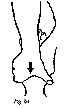
Figure 1a: thumb web adjustment.
Thumb web adjustment: With thumb webs of both hands contacting the contact point, doctor brings talus to tension by tractioning inferior and adding slight dorsiflexion. Doctor thrusts A-P and lateral-medial with triceps contraction.
Little finger contact adjustment: With both "little" fingers overlapped on the contact point and fingers laced (thumbs on sole of foot), doctor brings talus to tension with inferior traction and dorsiflexion. The thrust is a "scoop" in an A-P and lateral-medial direction.
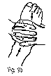
Figure 1b: little finger contact adjustment.
#6. Calcaneus - Everted and Plantarflexed Subluxation.
This bone may be adjusted/mobilized with the patient prone as shown, but the supine adjustment is preferred. Patient doesn't have to turn to the prone position from supine.
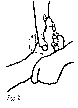
Figure 2: calcaneus adjustment.
Adjustment: The Doctor contacts the calcaneus with the palm of the "outside" hand. "Inside" hand "shakes hands" with the foot. The thenar of the "outside" hand applies slight lateral to medial pressure as "inside" hand performs several clockwise and counterclockwise rotations.
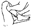
Figure 3: fibula adjustment.
#7. Fibula - Posterior and Lateral Subluxation.
The doctor will find point tenderness on the posterior aspect of the fibular head. This is the contact point.
Adjustment: The Doctor takes a tissue pull with metacarpal/interphalangeal joint of "outside" hand in an anterior and posterior and lateral to medial direction. "Inside" hand grasps patient's ankle and approximates the heel to the buttocks. The thrust is very quick, with the heel toward the buttocks (P-to-A).
#8. Cuboid - Superior and Lateral.
By far, the most common direction of cuboid subluxation. This subluxation is not corrected by an orthotic. Due to inversion sprains and ankle rolls, the cuboid appears to subluxate in a superior and lateral direction. Doctor will be able to see, and usually feel, this pattern.
Adjustment: The Doctor contacts the cuboid area on the lateral aspect of the foot with distal interphalangeal joint of "inside" thumb. "Outside" hand contacts directly over "inside" hand. With the doctor's hands on patient's foot, the foot is taken off the table laterally and placed between doctor's slightly bent knees.
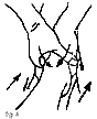
Figure 4: cuboid adjustment.
The thrust is achieved by doctor squeezing his hands and knees in a lateral to medial direction, along with extension of the knees. A characteristic "loud" audible will usually be heard.
(Part III, "Associated Pronation Adjustments," will appear in the June 4 issue.)
Mark Charrette,DC
Las Vegas, Nevada
Click here for previous articles by Mark Charrette, DC.