


We are exposed to radiation all the time. The average individual receives 3 mSv per year from natural sources including radioactive material and cosmic radiation from outer space. This is described as a "background" dose and varies throughout the world.
What is a millisievert (mSv)? It's the scientific unit of measurement for a radiation dose referred to as the effective dose. Because different tissues in the body have varying sensitivities to radiation exposure, the actual dose to different parts of the body will vary. Therefore, the term effective dose or sievert is used when referring to the dose averaged over the entire body. The effective dose allows for quantification of risk and comparison to sources of exposure from background radiation and radiographic medical procedures.
The X-ray examination provides valuable information and is crucial for accurate diagnoses. I am not suggesting X-ray examinations should be avoided, but I am suggesting we need to remember that ionizing radiation is a known carcinogen, and we need to keep that in mind when deciding which diagnostic tool to use.
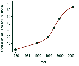 People employed in radiology before 1950 were exposed to high levels of radiation, mainly from fluoroscopy. They also had an increased mortality rate from cancer, usually leukemia. The first commercial computed-tomography (CT) scanner was developed in 1972. Since then, CT has become the major source of medical radiation. Conservative estimates reveal that more than 60 million CT examinations were done in 2002 in the U.S., representing an estimated 70 percent of all medical X-ray exposure.1 The use of CT has continued to increase, predominately because it represents perhaps the single mostimportant advance in diagnostic radiology.
However, as comparedwith plain-film radiography, CT involves much higher doses ofradiation, resulting in a marked increase in radiation exposurein the population. For example, a PA and lateral chest X-ray exposes the patient to 0.16 mSv, but one abdominal CT scan exposes the patient to 10.00 mSv. Plain-film radiography is not increasing radiation exposure in the population, but the use of CT is increasing, which directly increases the overall population's exposure to radiation. This is cause for concern.
| Approximate mean doses relevant to societal low-dose radiation exposures and to low-dose radiation risk estimation | Approximate mean individual dose (mSv)* |
| Round-trip flight, New York to London | 0.1 |
| Single screening mammogram (breast dose) | 3 |
| Background dose due to natural radiation exposure | 3/yr |
| Dose (over a 70-year period) to 0.5 million individuals in vicinity of Chernobyl accident | 14 |
| Dose range over 20-block radius from hypothetical nuclear terrorism incident | 3-30 |
| Pediatric CT scan (stomach dose from abdominal scan) | 25 |
| Radiation worker exposure limit | 20/yr |
| Exposure on International Space Station | 170/yr |
| A-bomb survivors [mean dose in LSS cohort] | 200 |
| Medical X-rays [breast dose in scoliosis study] | 100 |
| Nuclear workers [mean dose from major studies] | 20 |
| Individuals diagnostically exposed in utero | 10 |
| Source: Brenner DJ, Doll R, Goodhead DT, et al. Proc Natl Acad Sci USA, 2003;100:13761. | |
| Typical organ radiatation doses from various radiologic studies | ||
| Study Type | Relevant Organ | Relevant Organ Dose* (mGy or mSv) |
| Dental radiography | Brain | 0.005 |
| Posterior-anterior chest radiography | Lung | 0.01 |
| Lateral chest radiography | Lung | 0.15 |
| Screening mammography | Breast | 3 |
| Adult abdominal CT | Stomach | 10 |
| Barium enema | Colon | 15 |
| Neonatal abdominal CT | Stomach | 20 |
| *The radiation dose, a measure of ionizing energy absorbed per unit of mass, is expressed in grays (Gy) or milligrays (mGy); 1 Gy = 1 joule per kilogram. The radiation dose is often expressed as an equivalent dose in sieverts (Sv) or millisieverts (mSv). For x-ray radiation, which is the type used in CT scanners, 1 mSv = 1 mGy. Source: Brenner D, Hall E. NEJM, November 2007;357:2277-84. | ||
The quantitative information for radiation risks and radiation-induced cancers comes from studies involving survivors of the 1945 atomic bombing in Japan. This cohort of survivors has been intensely studied. The epidemiological study with thehighest statistical power for evaluating low-dose risks is theLSS cohort of atomic bomb survivors.2 Although these analysesoften have been considered high-dose studies, in fact, themean dose in the exposed group in the LSS cohort was only 200mSv, with more than 50 percent of the exposed individuals in the cohort (26,300individuals) having doses of less than 50 mSv. Cancer incidence,3 cancer mortality, and noncancer-related mortality havebeen studied, although almost half the exposed population (anda larger fraction of the individuals exposed as children) isstill alive.
In estimating the lowest dose of x- or gamma-radiation for evidenceof increased cancer risks, it is important to make the distinctionbetween acute exposures over a very short period (such as theatomic bomb exposures) and protracted exposures (such as occupationalor fractionated exposure). In general, protracted exposuresto x- or gamma-radiation are associated with lower risks than thoseof an acute exposure to the same total dose, both for cancer and other endpoints. There is another very difficult problem with estimating risks from lower doses of x- or gamma-radiation compared with higher doses. The risks are likely to be lower, and progressively larger epidemiologicalstudies are required to quantify the risk to a useful degreeof precision. For example, if the excess risk were proportional to the radiation dose, and if a sample size of 500 people was needed to quantify the effect of a 1,000-mSv dose, then a samplesize of 50,000 would be needed for a 100-mSv dose, and approximately 5 millionfor a 10-mSv dose. In other words, to maintain statisticalprecision and power, the necessary sample size increases approximatelyas the inverse square of the dose. As one can see, it is a difficult statistical project to evaluate such large populations.
The most complete information available presently regarding lower radiation exposure and cancer risks comes again from the LLS cohort of atomic bomb survivors exposed to less than 50 mSv with a mean dose of approximately 34 mSv, which approximates the relevant organ dose exposure from a typical CT study, in which two to three scans are done on a patient. There is no specific number or range to which scientists are willing or able to commit that would give us a guideline for when to be concerned about exposure risks.
One study involving more than 400,000 radiation workers in the nuclear industry has reported a significant association between radiation dose and mortality from cancer. Again, the range of exposure was between 5 and 150 mSv. Theses same risks were quantitatively consistent with those of the atomic bomb survivors.4
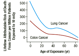 The risks also vary depending on the age of the patient at the time of exposure and the organ-specific dose exposure. Extrapolating from the data available, the risk for cancer-related death associated with one abdominal CT scan is 0.06 percent for a patient exposed at 25 years of age and 0.02 percent for a patient exposed at 50 years of age. This risk is remarkable when looking at the lifetime radiation risk of two of the most common radiogenic cancers: lung and colon. When exposed to just 10 mSv at 25 years of age, the risks for death from lung and colon cancer are 0.025 percent and 0.0125 percent, respectively. For a 50-year-old, these associated risks are 0.017 percent and 0.010 percent for lung and colon cancer, respectively.5
CT has revolutionized diagnosis and medical practice in the past 30 years, but clinicians and patients need to be aware of the associated risks, not just the clinical benefits. The most concerning issue is the growth of CT use on children, driven primarily bythe decrease in the time needed to perform a scan - nowless than 1 second. This largely eliminates the need foranesthesia to prevent the child from moving during image acquisition.The major growth area in CT use on children has been presurgicaldiagnosis of appendicitis, for which CT appears to be both accurateand cost-effective.
Another large part of the projected increase in CT scanning for adultscomes from new CT-based screening programs forasymptomatic patients. The four areas attracting the most interestare CT colonography (virtual colonoscopy), CT lung screeningfor current and former smokers,CT cardiac screeningand CT whole-body screening.These studies can be self-referrals, which makes it difficult to monitor if the patient is doing the referring. With self-referral, monitoring of the radiation dose becomes the patient's responsibility.
Clinicians and patients both need to be aware of the general exposures for the more common studies. The decision to have an X-ray exam is a medical one, based on the likelihood of benefit from the exam and the potential risk from radiation. For low-dose examinations (plain-film X-rays), the decision is not difficult; exposures are minimal therefore of very low risk. For higher-dose exams such as CT and barium studies and interventional radiology, the clinician needs to consider the patient's previous exposure and if there is another non-ionizing radiation test (MRI or ultrasound) that could give comparable information. Patients should keep a record of their X-ray history for themselves, especially CT scans, since their history may not be easily accessible for health care providers. Pregnant patients should avoid ionizing radiation examinations whenever possible.
How much is too much? At present, this question cannot be answered. Remember, the mean dose for the atomic bomb survivors was only 200mSv, with greater than 50 percent of the exposed individuals in the cohort having doses of less than 50 mSv. I suggest we keep in mind the lower-dose population when determining which test to order; particularly in cases in which the patient is young and has already had a CT scan. There is no right answer to the question as to how much is too much, but we should use the diagnostic tests that involve ionizing radiation with some consideration of lifetime exposure to radiation.
References
- Lee CI, Haims AH, Monico EP, et al. Radiology, 2004;231:393-8.
- Preston DL, Shimizu Y, Pierce DA, et al. Radiat Res, 2003;160:381-407.
- Pierce DA, Preston DL. Radiat Res, 2000;154:178-86.
- Cardis E, Vrijheld M, Bletter M, et al. Radiat Res, 2007;167:396-416.
- Brenner D, Hall E. NEJM ,2007; 357:2277-84.
Click here for more information about Deborah Pate, DC, DACBR.