


 Figure 1. This illustration shows the broken chair and the first point of contact with the floor. It also illustrates that shear strain would have resulted as the thoracic spine was abruptly decorated while the head�s inertia carried it rearward relative to the torso. The inset was used to explain the effect of shear on the discs that
were claimed to have been injured.
Currently, these tough answers emanate from a variety of experts including medical doctors, chiropractors, biomechanical engineers, and accident reconstructionists, to name the more common types. Based on my 27-year immersion in this field, I believe it is fair to state, with no intention of cynicism or hyperbole, that a substantial proportion of these so-experts are sorely lacking in one or more critical areas. (Of course, there are also some highly competent experts.) The opinions that are consequently tendered range from inexcusable ignorance to pure brilliance.
The essential nature of the problem, in my view, is in training or lack thereof. Accident reconstructions, for example, have little or no training in human biology and health. Biomechanists have some, but not enough, nor do they have clinical experience or the ability to read MRIs or interpret other medical documents or opinions. Most of them do not view this as a hindrance, of course, and very often the courts allow biomechanists quite a bit of latitude.
An appropriately trained medical or chiropractic doctor, on the other hand, can obtain the requisite skills to wear all of the hats necessary to put the entire case together so all of the crucial features of the case - accident history, biomechanical events, analysis of the effects of forces and loads on human tissues, and medical correlation or corroboration - can be linked. Because there are not many of these "appropriately trained" hybrids around, the field is quite open and in need of people to fill the vacuum.
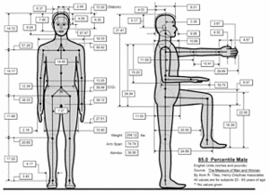 Figure 2. This image represents the mean dimensions
of a man of the patient's height, an 85th percentile male. The height of the torso of a seated man who is 6 ft tall is 43.33 inches. [Contact Dr. Croft at
![]() for specific data from this image.]
Let's take a look at a recent case I worked on. I chose this case as an example because (a) It is not a whiplash case, per se; (b) Although it has medical and humanitarian merit, from a legal standpoint it would be considered challenging by most lawyers; and (c) It illustrates the all-important A-to-Z linking, making connections from cause to effect, and the critical medical corroboration.
for specific data from this image.]
Let's take a look at a recent case I worked on. I chose this case as an example because (a) It is not a whiplash case, per se; (b) Although it has medical and humanitarian merit, from a legal standpoint it would be considered challenging by most lawyers; and (c) It illustrates the all-important A-to-Z linking, making connections from cause to effect, and the critical medical corroboration.
The Case
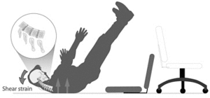
A middle-aged man was sitting in an office chair. When he leaned back slightly, the chair back broke and the man fell off the chair, landing on his thoracic spine with his legs in the air (Figure 1). (Note: I made these illustrations for two posters that were used at the man's settlement conference.)
Figure 1. This illustration shows the broken chair and the first point of contact with the floor. It also illustrates that shear strain would have resulted as the thoracic spine was abruptly decorated while the head's inertia carried it rearward relative to the torso. The inset was used to explain the effect of shear on the discs that were claimed to have been injured.
 From the figure, it is clear that we can actually equate this fall with a whiplash phenomenon. This is convenient because we have a lot of special knowledge concerning the forces and mechanics of this form of injury. But first we need to understand, quantitatively, the severity of the impact. Using a program called AnthroCalc, I determined the approximate dimensions of various body parts and their height above the floor before the fall (Figure 2). From the dimensions determined, the approximate fall velocity can be calculated as follows, using simple Newtonian principles:
In this equation Ve represents ending velocity in feet per second (fps); Vi represents initial velocity at the start of the fall, which, in this case, is 0 fps; a represents acceleration due to Earth's gravity (32.2 fps2); and d is the distance of the fall in feet (43.3 inches is 3.6 feet). The average acceleration can be derived as well:
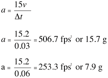
In this equation, delta V is the change in velocity, obtained from the first equation, and delta t is the actual duration of the change of velocity. In the first equation there is not much uncertainty, since we have the actual chair and can approximate, with reasonable accuracy, the fall height. I could have considered some uncertainty there, but decided it would have been trivial. The duration, delta t, on the other hand, cannot be precisely estimated, so I used two values, 0.03 seconds and 0.06 seconds, as bracketed ranges of uncertainty there. Dividing the derived acceleration values by 32.2 fps2 takes us back to the more familiar gravitational acceleration term, g.
So, now we have a range of probable acceleration from 7.9 to 15.7 g. This is torso acceleration. The head's acceleration would have been higher still. It is important now to anchor these figures to something tangible. Experimental studies using cadaver spines show C4-5 and C5-6 are at risk for disc rupture at 3.5 g, and other levels at higher accelerations of 5 g and higher.1 Therefore, the probability of disc rupture in this fall is high.
A complicating feature of this case was the man's long history with neck pain. Only several weeks before his fall, an MRI had revealed three cervical disc herniations and rather significant multi-level spondylosis. He had also been treated with a cervical epidural steroid injection shortly before the fall. When I examined the MRI taken after the chair accident, I found that the cervical herniations had increased in their horizontal dimension. However, because the two studies had dissimilar magnifications, and because there was no way to calibrate dimension on either study, actual measurements in millimeters could not be compared.
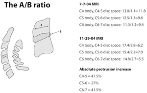 Figure 3. The first MRI (7-7-04) was obtained prior to the fall and the second MRI (11-29-04) was obtained after the fall. The absolute or A/B ratio can be obtained even though the two studies were not calibrated in such a way as to allow direct measurement.
Instead, I simply compared the measured A/B ratios obtained at the affected levels on the two MRI studies (Figure 3). Since these are simply ratios, it is not necessary to know what the unit dimensions are. I found that the three herniations had increased by 47.5 percent, 21.5 percent and 41.4 percent at levels C4-5, C5-6, and C6-7, respectively. Moreover, type I Modic changes were visible only on the second MRI. These changes are indicative of an inflammatory reaction. The man ultimately underwent a tri-level fusion with plates and screws.
Figure 3. The first MRI (7-7-04) was obtained prior to the fall and the second MRI (11-29-04) was obtained after the fall. The absolute or A/B ratio can be obtained even though the two studies were not calibrated in such a way as to allow direct measurement.
Instead, I simply compared the measured A/B ratios obtained at the affected levels on the two MRI studies (Figure 3). Since these are simply ratios, it is not necessary to know what the unit dimensions are. I found that the three herniations had increased by 47.5 percent, 21.5 percent and 41.4 percent at levels C4-5, C5-6, and C6-7, respectively. Moreover, type I Modic changes were visible only on the second MRI. These changes are indicative of an inflammatory reaction. The man ultimately underwent a tri-level fusion with plates and screws.
As challenging as this case might have appeared, it actually settled for well into the six-figure range and did not go to trial. It is tempting to speculate that my analysis was a critical determinant in this outcome, but when a case is realistically "reconstructed" in this way, it has been my experience that the parties are more likely to reach a mutually acceptable neutral ground short of an actual trial. In this example, it should be clear that a non-physician biomechanist would not have been able to read and interpret the MRIs - a critical link between the mechanism and the outcome. The orthopaedic surgeon did not attempt to compare the MRIs critically or quantitatively, nor was he able to provide the biomechanical commentary on forces or their relation to disc injury. These unlinked and unidentified features would have made the case much more difficult if they had not been revealed.
Reference
- Panjabi MM, Ito S, Pearson AM, Ivancic PC. Injury mechanisms of the cervical intervertebral disc during simulated whiplash. Spine, June 1, 2004;29(11):1217-25.
Click here for previous articles by Arthur Croft, DC, MS, MPH, FACO.