


Editor's note: This is the second in a series of three articles on SI mobilization. The first article appeared in the Nov. 4 issue.
I have a memory, now at least 32 years old, of Dr. Hogan, my technique teacher at National. I was a "know it all" back then, but for some reason I paid attention when he said: "Adjust the sacrum, not the ilium." I think he was referring to the tendency of the ilial side of the joint to compensate, and the tendency of practitioners to adjust the easily assessed iliosacral joint. You often have to address the sacral side of the joint separately.
I have gone back and forth with using the muscle energy model of the sacroiliac (an osteopathic low-force spinal adjusting technique using post-isometric relaxation) for the past 16 years. In my opinion, the model is a bit cumbersome, the terminology has never really made sense to me, and the palpation system is nothing like the rest of the motion palpation that I use - but despite all these drawbacks, muscle energy seems to address the sacrum more effectively than anything else I have used. Once I started using the stork test again, I realized how many sacral fixations I was missing, and that my previous corrections didn't always correct the fixation. I am once again using this model daily.
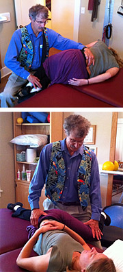 Top: Position for correction of left anterior sacral torsion; Bottom: Position for correction of left posterior sacral torsion.
I will repeat what I said in the previous article about the sacral side of the SI joint. In the stork or Gillet test, when you are testing the weight-bearing side, you are testing how well the sacrum is moving on the ilium. When the patient lifts their bent right leg as high as they can, we are testing the left-side sacral function. Your left thumb is on the left PSIS and your right thumb is just medial to this, on the sacrum, at the left sacral base at S2 or so. Can your right thumb, on the left upper sacrum, appear to drop inferior as the right bent leg is lifted to end range? If your thumbs appear to move inferior together, the left sacrum is fixated.
Top: Position for correction of left anterior sacral torsion; Bottom: Position for correction of left posterior sacral torsion.
I will repeat what I said in the previous article about the sacral side of the SI joint. In the stork or Gillet test, when you are testing the weight-bearing side, you are testing how well the sacrum is moving on the ilium. When the patient lifts their bent right leg as high as they can, we are testing the left-side sacral function. Your left thumb is on the left PSIS and your right thumb is just medial to this, on the sacrum, at the left sacral base at S2 or so. Can your right thumb, on the left upper sacrum, appear to drop inferior as the right bent leg is lifted to end range? If your thumbs appear to move inferior together, the left sacrum is fixated.
The next part, to a chiropractor, will feel like a weird variation on motion palpation. Instead of moving a bone, you are assessing as the patient moves around your hands. Much of this assessment is visual, rather than kinesthetic.
The patient lies prone. If the left sacrum is stuck, I am first assessing the depth of the sacral sulcus, at the sacral base, comparing one side to the other. Using your dominant eye, look directly in the midline, looking down at your thumbs on the sacrum. This is both a visual and a positional palpation finding. In anterior or posterior torsions, the involved side will be more superficial, so the opposite side looks deep. You will also assess the horizontal level at what the osteopaths call the ILA, the inferior lateral angle near the bottom of the sacrum.
Line up your fingers on what feels like the same structure on each side on the lower sacrum. I like to do this twice, once coming more posterior to anterior on the sides of the sacrum, and the other coming up from below. This one is a pretty pure visual test. In either of these torsions, the ILA will be inferior or more prominent (as if it is rotated back) on the fixated side.
What does this osteopathic terminology, sacral torsion, mean? It implies that the sacrum is twisted around an oblique axis. In both cases, both in a left anterior torsion (called left on left; don't even ask me what that means), and in a left posterior torsion (called left on right); the left sacral base palpates and visualizes as posterior. The left ILA is more prominent in both of these lesions. The anterior torsion is considered more of a compensation, as if the gait got stuck at one end of the motion. A descriptive term for this might be sacrum vulgaris or the common sacral dysfunction. It is sometimes described as a problem with piriformis hypertonicity.
The posterior torsion, or stuck posterior pattern, is a more significant fixation, completely restricting any extension motion of the lower lumbar and sacral spine. When you have a posterior torsion, you will often also find a lower lumbar vertebral joint that resists extension and side-bending. A posterior sacral torsion completely locks up the lower back. If it occurs concurrent with an anteriorly rotated ilium, it can be even more problematic.
These patients may present with sciatica, which may be coming from the stuck SI joints rather than from a true disc lesion. Or perhaps the stuck sacrum and lumbars are putting further stress on an already compromised disc. They may not get relief from McKenzie extension, as they cannot extend with ease. Once you correct both the posterior sacral torsion, and the stuck-in-extension lumbar vertebrae, then self-mobilization into extension is likely to be helpful.
Differentiating an Anterior Torsion From a Posterior Torsion
The static palpation findings are the same for an anterior and a posterior torsion. With both, the sacrum is more superficial, more prominent on the fixated side. With both, the ILA is more prominent on the fixated side.
We differentiate them with motion. In the muscle energy model, we use the patient's own motion. The patient lies prone; your thumbs are in the bilateral sacral sulcus. In our example, the left sacral sulcus is more prominent. Now, ask the patient to prop themselves up on their elbows or push up with their arms, leaving the pelvis on the table. They are producing extension in the lower spine. You are watching and feeling what happens at the sacral sulcus. If this extension motion helps correct the sacral unleveling, you are looking at an anterior torsion. If the extension motion either leaves the sacral unleveling unchanged or makes it worse, you are looking at a posterior torsion. As I mentioned, in a posterior sacral torsion, the lumbosacral junction loses all of its ability to extend. You can just spring the lumbosacral junction here; it will be very rigid in a posterior torsion.
Correcting the Sacral Torsions
In the muscle energy model, for any fixation, you first take the patient to the soft end-range in all three directions. You then use post-isometric relaxation, having the patient contract and then relax. As they relax, the fixated joint moves farther into the barrier, freeing up the joint. I suspect that this is the aspect of muscle energy that is least well understood. The trick to doing a good job with the osteopathic styles of low-force adjusting is in the subtlety of finding the end play, and of positioning the patient at the soft end-range of the barrier. It's like a good paint job: all the hard work is in the preparation. There are variations in how these techniques are done. A master of muscle energy makes it look easy, setting it up so the body just seems to correct itself.
Here is how to correct an anterior torsion. The goal is to release the anterior (deep) side - the right side in our example - back into place. In our example, the left side is both fixated and more superficial, so the lesion is a left anterior torsion. The patient lies on their left side with the left arm behind them. Having the left arm behind twists the upper body farther into a left rotation.
Move the patient's right arm and upper body to the end range of that rotation. Bring both of the patient's bent legs off the table, and bring the lumbar spine farther into flexion. As you do this, you palpate the lumbosacral joint and take the legs into enough flexion to begin to open the lumbosacral joint.
Now, have the patient push in a superior direction - toward the ceiling - with both feet for a five count, against your resistance; when they relax, take up the slack on all of the barriers. Repeat two more times. If you have never done this style of muscle energy, you will be surprised by how the barrier seems to soften and recede during this seemingly minimal procedure. Your right hand is monitoring at the lumbosacral junction, but not really doing the correction.
The usual instruction is for the patient to push very gently; most patients will try to push hard. I like to sit on a stool. You are supporting both of the patient's knees on your thighs. I often use a small pillow on my thighs, just for my own comfort. Remember, you are releasing the deep side. As the patient is left-side lying, your focus, at least for a typical chiropractic move, would be on the right side of the involved sacrum. There is no thrust in any muscle energy adjustments. It's all about the positioning and the contract-relax to reset the joint. The hardest part for a typical chiropractor is the absence of pushing or thrusting; just let the correction happen.
Here is how to correct a posterior torsion. I've seen several variations on this move. (There is a truly elegant YouTube video of another variation on this correction - which the presenter calls a left on right, the osteopathic terminology for the same posterior torsion lesion - at www.youtube.com/watch?v=_tYiv4cSzbs&feature=related. The correction I commonly use for a left posterior torsion involves the patient right-side lying with the left side up, both arms in front. It starts out like a typical chiropractic side-posture move.
Roll the patient's left shoulder back, taking out the slack of the left rotation. Then bring the bent patient's upper leg into about 90 degrees of flexion. Make sure the patient's lower leg stays straight and move it slightly backward into slight extension on the table. The fingers of my right hand are monitoring the left sacrum, but not really actively doing any correction.
The contract relax can be done in two ways in these techniques. In the first version, the patient pushes their bent knee upward toward the ceiling against the isometric resistance of your left hand for a five count. They then relax, and you take up the slack on all of the barriers, especially focused on rotation, bringing the bent leg toward the floor. Repeat two times. Another version uses a scissors type of movement. In this one, the patient isometrically extends or straightens the bent left leg against your left thigh's resistance, and flexes the straight right leg against your right thigh. As they relax, you move the lower leg farther into extension and the upper leg farther into flexion. In either version, you'll feel the left side of the sacrum release.
I know that the written word is not always the ideal way to teach technique. At www.youtube.com/marchellerdc, I've posted videos of these two techniques. I also know one article is not enough to learn a new model or new technique. Some of you may start using the Gillet/stork test more consistently, and finding out whether your typical sacral techniques are working to release the sacrum. Others may want to further pursue a deeper understanding of muscle energy methods.
Resources
- Comerford M. Mobilization of the Sacroiliac Joint (class), June 2008, Kinetic Control.
- Whyte-Ferguson L. Clinical Mastery in the Treatment of Myofascial Pain. Lippincott, Williams and Wilkins, 2005.
- Greenman's Principles of Manual Medicine, 4th edition. Lippincott, Williams and Wilkins, 2011. An earlier edition of this was my bible for muscle energy technique
Click here for more information about Marc Heller, DC.