


One of the many joys I experience in treating patients is when I can address areas of the body that are not run of the mill for chiropractors. Although I love treating the spine, extremity problems are exciting and it's the "thrill of the hunt" that keeps me enthused.
Think for a moment about how many of your patients complain of foot pain. After 16 years in practice and treating lots of extremity pain and athletes, I estimate about 10 percent of my patients present with this complaint at one time or another.
If extremities aren't your area of expertise, side-stepping this issue and pointing out faulty shoes while yanking on the patient's heel may seem like enough attention. It isn't enough. I want to help improve your clinical skills; to do that, let's start with a quick anatomy review.
Heel Anatomy
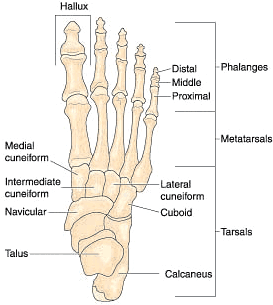
The heel itself is the prominence at the posterior end of the foot. It is based on the projection of one bone, the calcaneus or heel bone, behind and underneath the articulation of the ankle mortise (tibia and talus).
The compressive forces applied to the foot during weight-bearing activities are distributed along the five toes and metatarsals; three on the medial side and two on the lateral. The lateral support stretches over the cuboid to the heel and the medial support moves over the three cuneiform bones and the navicular to the heel.
These bones all help form the three functional arches of the foot (medial longitudinal, lateral longitudinal, transverse) that are optimized to distribute compressive forces across an uneven terrain. Due to the anatomy, the heel forms the posterior point of support. Along with the ball of the large toe (1st MT) and little toe (5th MT), they bear the brunt of the loads from heel strike and standing.
 In an attempt to distribute the compressive forces placed on the heel during weight-bearing activities, especially when the heel contacts the ground during gait, the sole of the foot is covered by a layer of subcutaneous connective tissue. It can get as thick as 2 cm under the heel. This tissue has a system of pressure chambers that act to absorb shock and stabilize the sole. Each of these chambers contains fibrous fatty tissue covered by a layer of tough connective tissue made of collagen fibers. These tissues are firmly attached both to the plantar aponeurosis (plantar fascia) above and the sole's skin below. The sole of the foot is one of the most highly vascularized regions of the body.
The Achilles tendon is the muscle tendon of the triceps surae "three-headed" group of muscles that includes the soleus and the two heads of the gastrocnemius. The main function of the triceps surae is plantar flexion. The plantaris muscle also attaches to the heel bone, but it is not visible.
The key idea to keep in mind is that the heel ends up being the actual point of contact during the heel-strike portion of the gait cycle. It is also an important point on which weight is equally balanced from the forefoot and heel. We need 50/50 weight distribution at the forefoot and hindfoot to have healthy walking and standing.
When all things are healthy and the arches are working well during gait, our heel strikes the ground in a slightly supinated position and moves into slight pronation during foot flat. Stand up and walk a few paces; notice how it feels. Based on my clinical experience, healthy gait happens only in about 10 percent of patients.
The more likely scenario is to have a patient who is overpronating (in my experience, at least 80 percent of patients). Since the foot is now flattening and the arches are collapsing more into excessive pronation, this causes the heel to strike the ground in an oversupinated position. This altered heel-strike pattern creates not only stress in the heel, but also altered biomechanical patterns that can lead to pain. Excessive supination occurs much less frequently (most statistics suggest less than 5 percent of the population), so its effects on the heel are not as profound.
Common Conditions
Achilles Tendonitis: The tendon of the triceps surae falls prey to excessive pronation of the feet. The downward collapse of the foot creates tension on the Achilles tendon, causing it to bow inward. This inward bowing is happening every time the overpronating patient is weight-bearing. It creates microtrauma and stress to the Achilles tendon. For supinators, the bowing of the Achilles tendons is outward. Similar strain and stress on the bones and soft tissue may occur as well.
Over time, spurs can develop on the posterior, superior portion of the calcaneus bone. Hypertonicity and trigger points in the gastrocnemius and soleus muscles occur. Achilles tendonitis can be debilitating because the pain will continue to alter gait and mobility patterns to the point that it prohibits walking any appreciable distance without pain.
Conservative care has been shown to yield positive results to patients with Achilles tendinopathy. "A combination of conservative rehabilitation strategies may be used by chiropractors to treat midportion Achilles tendinopathy and allow an individual to return to pain-free activities of daily living (ADLs) in a timely manner."2
Plantar Fasciitis: When the foot is healthy and the arches are working well, the plantar fascia is a healthy ligament that exhibits elastic properties. Over time, the three arches of the foot tend to collapse to the floor. This excessive pronation stresses the plantar fascia chronically, causing it to stay overstretched and lose its elasticity. It becomes longer due to the plastic deformation.
Supination creates a thinner, tighter foot due to the tendency of the foot to roll outward. These patients have a high instep with collapsed transverse and lateral arches.
In either case, we can note biomechanical instability and irritation to the plantar fascia from its origin at the calcaneal tubercle across to the insertion on the metatarsal heads. Generally, symptoms begin as a dull ache in the underside of the heel that is more of an annoyance. Patients can still perform normal activities and sports.
However, as time goes on and the pain becomes more intense and sharp, they have to curtail or eventually stop their activities. In the later phases, pain can move off the heel and into the middle of the fascia, or at the insertion on the metatarsal heads.
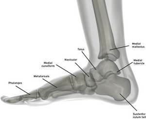 Sprained Ankle:: Ankle sprains range from the very mild kind to the severe. The milder types are suggested when a patient complains of "turning the ankle." This is like when someone missteps off a curb or a step. The opposite extreme is when ligaments and tendons are torn due to a traumatic injury. Either way, biomechanics, particularly of the calcaneus and talus, are compromised.
On a typical inversion ankle sprain, the calcaneus bone moves medial while the talus tends to move lateral. In the event of an eversion sprain, the opposite bony movement occurs. Recall that the bony anatomy allows inversion ankle sprains to occur much more frequently. In either case, the movement of the bones inhibits normal mobility. Since most people do not see a chiropractor after a sprained ankle, the bones stay out of alignment, leading to a slow rate of healing, chronic degree of tenderness and decreased function, and a tendency to repeatedly sprain the ankle in the future. Thus, it is imperative that chiropractors intervene and break this cycle.
To help avoid frequent recurrence of ankle sprains, a 2004 study suggested "high velocity, low amplitude chiropractic manipulative therapy to the spine, pelvis, and extremities, particularly at the ankle, should be considered when managing young recreational athletes with functional chronic, recurrent, ankle inversion sprains."3
Heel Spurs: As previously discussed, a heel spur forms at the posterior, superior calcaneus bone courtesy of Achilles tendonitis. The most common place we see a spur is on the underside of the foot at the calcaneal tubercle where the plantar fascia originates. The worse the chronic pull on the bone, the worse the spur.
We can't take the spur away, of course, but we can help it from getting worse. Depending on how quickly you are able to get to the patient with one or more of the above ailments, the faster they heal. If a patient is acute enough and there is sharp pain and inflammation, modalities like cold laser, ultrasound, ice, etc., are indicated. You can also provide light, soft-tissue therapy at this stage, making sure you apply the appropriate amount of pressure.
Treatment Tips
Adjusting the bones of the feet to restore healthy biomechanics is extremely important. Adjusting the calcaneus, talus, navicular and cuboid bones is particularly helpful. If you don't get in there and move those bones, they will not heal very well and will be prone to future injuries.
Use your head on this. If the person is in a lot of pain, start with light-force techniques, like a spring-loaded instrument. Then proceed to light manual work with your hands until you can adjust the patient fully and properly. So many doctors skip adjusting the foot, but as you can tell, it's an important part of your whole-body care.
Elastic taping, especially in the initial stages of care, will help support the tissues, and promote good circulation and movement patterns. Generally, a "figure 8" tape job works well here. You can move the foot into dorsiflexion and plantarflexion while taping to promote as full a range of motion as possible. Taping ankles for the purpose of treating Achilles tendinitis has shown an increase in the active ankle joint range of motion, and a decrease in tenderness and pain.4
Stabilizing orthotics that are flexible, have all three arches and are custom molded will greatly support the patient's feet and the rest of their body. A 2002 study suggests the effectiveness of applying orthotics and ankle braces during the acute and subacute phases of ankle rehabilitation.5 The use of orthotics, when necessary, has been a part of my practice for 16 years and in my experience, they do a phenomenal job helping maintain proper biomechanics and helping adjustments hold longer.
Ankle stretches and stabilization exercises also can be provided, and they are quite easy. Have the patient cross the affected ankle over the opposite leg and passively dorsiflex, plantarflex, invert and evert the foot. Have the patient stand on the edge of a step or a door frame and stretch the calf muscles. Instruct them to hold the stretches described above in each direction for 30 seconds.
The stabilization exercises should be performed with elastic tubing, moving into dorsiflexion / plantarflexion / eversion and inversion. Elastic tubing is easy and convenient, and you can change the resistance quickly.
The ankle region is a common area of complaint from our patients as they walk into our offices. If we take a few moments to listen, look and feel, we can help them, and in a manner that isn't complicated or time consuming. Since the ankle is such an important stability structure for the entire body, our patients stand to benefit tremendously from our expertise in caring for them.
References
- Daniels CJ, Morrell AP. Chiropractic management of pediatric plantar fasciitis: a case report. J Chiropr Med, 2012 Mar;11(1):58-63.
- Papa JA. Conservative management of Achilles tendinopathy: a case report. J Can Chiropr Assoc, 2012 Sep;56(3):216-24.
- Gillman SF. The impact of chiropractic manipulative therapy on chronic recurrent lateral ankle sprain syndrome in two young athletes. J Chiropr Med, 2004 Autumn;3(4):153-9.
- Lee JH, Yoo WG. Treatment of chronic Achilles tendon pain by Kinesio taping in an amateur badminton player. Phys Ther Sport, 2012 May;13(2):115-9.
- Mattacola CG, Dwyer MK. Rehabilitation of the ankle after acute sprain or chronic instability. J Athl Train, 2002 Dec;37(4):413-429.
Dr. Kevin Wong, earned a BS in exercise physiology from the University of California – Davis and his DC degree from Palmer Chiropractic College West. He practices in Orinda, Calif., and serves the Lamorinda, Berkeley, Walnut Creek and many other East San Francisco Bay Area communities. He is an expert on foot analysis, walking and standing postures, and orthotics, and lectures nationwide on spinal and extremity adjusting.