


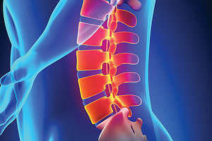
Lumbar facet denervation, more appropriately termed medial branch neurotomy (MBN), is a procedure that may be considered when patients suffer from recalcitrant non-radicular axial back and/or leg pain.
- How reliably can lumbar facet pain be diagnosed?
- What results can be expected from the procedure?
- What are the risks associated with the procedure?
- Will this procedure offer benefit beyond what has already been attained?
Pain in Facet Joints: Tough to Track Down
It has long been known and is now well-accepted that the facet joints are structures capable of producing pain. In 1963, Hirsh, et al., first demonstrated the capacity of the facet joint to produce pain by the injection of hypertonic saline into the joint.1 Mooney and Robertson later delineated their typical lower extremity referral patterns now well-known to most of us.2 Subsequent investigators have further confirmed the presence of facet joint nociceptive fibers capable of pain transmission.3-5
 However, less well-agreed-upon, compared to the joint's capacity for pain production, is the frequency with which this occurs and the precision with which it can be accurately diagnosed. The reported incidence of lumbar facetogenic pain shows wide variation. Most studies note an occurrence between 15-45 percent, depending on the diagnostic criteria utilized.6-8 Schwartzer, et al., have even suggested a 4 percent rate for the facet joint serving as a sole source of pain.9
Diagnostic precision for implicating facet pain is compromised in ways similar to that of confirming other potential sources of structural involvement. Spinal imaging, in general, is of little value in most non-neurogenic back pain syndromes.10-11 Pertaining specifically to the facet joints, abnormal imaging changes have been shown to have no relationship to the presence or magnitude of pain experienced.12-14
Adding to the diagnostic dilemma is that no particular pain pattern, clinical bedside test or combination of tests can consistently and reliably distinguish facet-mediated pain from that originating in other structures.15-17
Diagnostic Considerations: Pros & Cons of Nerve Blocks
In 1998, Revel, et al., demonstrated that patients matching 5/7 clinical criteria exhibited a 75 percent pain reduction on subsequent anesthetic blocks. These criteria were: age > 65, pain well-relieved by recumbent position, absence of pain with coughing, pain not worsened by forward flexion, pain not worsened when rising from flexion, pain not worsened by hyperextension, and pain not worsened by extension-rotation test.18 Further studies have shown that matching these criteria provides high specificity, but very low sensitivity in identifying those likely to benefit from anesthetic blocks.19-20
In the absence of predictive clinical or radiologic findings, nerve blocks are considered to be the best way of diagnosing presumed facet-mediated pain.8,21-22 However, at present, there is no clear consensus on how a diagnostic block should be performed, or the threshold and duration of pain relief that constitutes a positive response. This is largely due to the lack of a gold standard of diagnosis to which nerve blocks could be compared.21
Controlled diagnostic blocks imply having a patient undergo two separate injections, at different times, using anesthetic agents of different durations of action. A positive response occurs when a threshold of pain relief (usually between 50-80 percent) is experienced and the duration of relief is consistent with the known duration of the anesthetic. Single diagnostic blocks use only a single injection and anesthetic agent.
There are pros and cons to each approach. Falco, et al., in what appears to be the most recent review on the accuracy of these procedures, found that false positive rates ranged between 17-66 percent, being more common when single blocks or thresholds of pain relief less than 75 percent were used.8 Derby, et al., confirmed this finding, also noting that using pain-relief thresholds above 75 percent correlates with improved outcomes after medial branch neurotomy.23
However, additional research by Derby illustrated that using such highly specific criteria may unnecessarily exclude patients who could potentially benefit from interventional denervation. He found that 20 percent of patients with less than 50 percent relief, and up to 47 percent of patients with 50-69 percent relief (each considered to be negative responders to diagnostic blocks), went on to show a greater than 50 percent improvement in pain after undergoing medial branch ablation.24
It is also important to understand that pain relief subsequent to medial branch blocks does not implicate the facet joint with certainty. The medial branch also provides innervation to the multifidus, interspinous ligaments, periosteum of the neural arch, and the interspinal muscles.25-26 Ackerman, et al., further illustrated how the facet joints can be falsely implicated by demonstrating that anesthetic infiltration of the paraspinals, without direct anesthesia of the medial branch nerve, can lead to similar reductions in axial low back pain.27
Medial Branch Neurotomy: Let's Look at the Research
Although medial branch neurotomy may benefit properly selected patients, the relief achieved is rarely complete or permanent. Because of this, treatment decisions are best based upon having a realistic understanding of expected outcomes in relation to a patient's current level of pain and physical function.
Dreyfuss, et al., followed 15 patients showing >80 percent relief on controlled diagnostic blocks. Thirteen had relief of >60 percent at one year, with nine exceeding 90 percent pain reduction.28 Lakemeir, et al., assessed the six-month response to medial branch neurotomy in 29 patients after showing a minimum of 50 percent pain relief to a single diagnostic block. Average pain scale reduced from 6.6 to 4.7. Oswestry Index reduced from 40.8 to 28. This study also compared facet denervation to intra-articular steroid injection, finding no statistical difference between the two procedures.29
The response to radiofrequency rhizotomy after having successful comparative nerve blocks in Goldfeld, et al.'s study of 174 patients showed 119 having good (50 percent) to excellent (80 percent) pain relief and 55 showing no improvement. Ninety-six of those with good-excellent responses had relief lasting between 6-24 months, with 43 percent of that cohort showing sustained benefit for two years.30
Cohen, et al., followed 262 patients who had a positive controlled diagnostic block with >50 percent pain relief. Following medial branch neurotomy, 54 percent had pain relief >50 percent lasting at least six months. There was no difference in response between those reporting >80 percent relief on confirmatory blocks as compared to those reporting relief of between 50-80 percent.31
A later Cohen study reinforced this finding, further concluding that the use of more stringent diagnostic criteria (higher pain-relief thresholds or double as compared to single blocks) would likely result in withholding a beneficial procedure from a substantial number of patients without a corresponding improvement in success rates.32
Not all studies have shown favorable results for medial branch neurotomy. One of the largest double-blind, randomized trials found no difference in VAS scores, physical function or medication use between active intervention and sham groups.33 Leclair, et al., found similar disappointing results after monitoring 70 patients who had experienced relief after a single diagnostic facet injection. At 12 weeks, no difference was seen between an active treatment and sham group.34
Although this was one of the larger studies on facet denervation, it has been criticized for the lack of an adequate description of what constituted a positive diagnostic block.35 Similar criticism of reports finding evidence of procedural ineffectiveness have been echoed by Bogduk, et al., who stated: "Negative results have been reported only in studies that selected inappropriate patients or used surgically inaccurate techniques."36
Editor's Note: Part 2 of this article will continue the discussion of the benefits / risks of medial branch neurotomy, including key points for chiropractors to understand and share with patients who may be considering / advised to undergo this procedure.
References
- Hirsch C, Ingelmark BE, Miller M. The anatomical basis for low back pain. Studies on the presence of sensory nerve endings in ligamentous, capsular and intervertebral disc structures in the human lumbar spine. Acta Orthop Scand, 1963;33:1-17.
- Mooney V, Robertson J. The facet syndrome. Clin Orthop Relat Res, Mar-Apr 1976;115:149-56.
- Cavanaugh JM, Lu Y, Chen C, Kallakuri S. Pain generation in lumbar and cervical facet joints. J Bone Joint Surg (Am), 2006;88:63-67.
- Bogduk N, Wilson AS, Tynan W. The human lumbar dorsal rami. J Anat, 1982;134:383-397.
- Cavanaugh JM, Ozaktay AC, Yamashita T, et al. Mechanisms of low back pain: a neurophysiologic and neuroanatomic study. Clin Orthop Relat Res, 1997;335:166-180.
- Manchikanti L, Boswell MV, Singh V, et al. Prevalence of facet joint pain in chronic spinal pain of cervical, thoracic, and lumbar regions. BMC Musculoskelet Disord, 2004;5:15.
- Manchikanti L, Singh V, Pampati V, et al. Evaluation of the relative contributions of various structures in chronic low back pain. Pain Physician, 2001;4:308-316.
- Falco FJ1, Manchikanti L, Datta S, et al. An update of the systematic assessment of the diagnostic accuracy of lumbar facet joint nerve blocks. Pain Physician, 2012 Nov-Dec;15(6).
- Schwarzer AC, Aprill C, Derby R, et al. Clinical features of patients with pain stemming from the lumbar zygapophysial joints. Is the lumbar facet syndrome a clinical entity? Spine, 1994;19:1132-1137.
- Bechara BP1, Agarwal V, Boardman J, et al. Correlation of pain with objective quantification of magnetic resonance images in older adults with chronic low back pain. Spine, 2014 Mar 15;39(6):469-75.
- Boden SD, et al. Abnormal magnetic resonance scans of the lumbar spine in asymptomatic subjects: a prospective investigation. J Bone Joint Surg (Am), 1990;72A:403-408.
- Makki D, Khazim R, Zaidan AA, et al. Single photon emission computerized tomography (SPECT) scan-positive facet joints and other spinal structures in a hospital wide population with spinal pain. Spine J, 2010;10:58-62.
- Simon P, et al. In vivo topographic analysis of lumbar facet joint space width distribution in healthy and symptomatic subjects. Spine, 2012;37(12):1058-64.
- Kalichman L, Li L, Kim DH, et al. Facet joint osteoarthritis and low back pain in the community-based population. Spine, 2008;33:2560-65.
- Schwarzer AC, Aprill C, Derby R, Fortin JD, Kine G, Bogduk N. Clinical features of patients with pain stemming from the lumbar zygapophysial joints. Is the lumbar facet syndrome a clinical entity? Spine, 1994;15:1132-1137.
- Manchikanti L, Pampati V, Fellows B, Baha GA. The inability of the clinical picture to characterize pain from facet joints. Pain Physician, 2000;3:158-166.
- Hancock MJ, Maher CG, Latimer L, et al. Systematic review of tests to identify the disc, SIJ or facet joint as the source of low back pain. Eur Spine J, Oct 2007;16(10):1539-1550.
- Revel M, Poiraudeau S, Auleley GR, et al. Capacity of the clinical picture to characterize low back pain relieved by facet joint anesthesia. Proposed criteria to identify patients with painful facet joints. Spine, 1998;23(18):1972-6.
- Hancock MJ, et al. Op Cit.
- Laslett M, Öberg B, Aprill CN, McDonald B. Zygapophysial joint blocks in chronic low back pain: a test of Revel's model as a screening test. BMC Musculoskelet Disord, 2004;5:43.
- Sehgal N, Dunbar EE, Shah RV, Colson J. Systematic review of diagnostic utility of facet (zygapophysial) joint injections in chronic spinal pain: an update. Pain Physician, 2007;10(1):213-228.
- Binder DS, Nampiaparampil DE. The provocative lumbar facet joint. Curr Rev Musculoskelet Med, Mar 2009;2(1):15-24.
- Derby R, Melnik I, Lee JE, Lee SH. Correlation of lumbar medial branch neurotomy results with diagnostic medial branch block cutoff values to optimize therapeutic outcome. Pain Med, 2012 Dec;13(12)1533-46.
- Derby R, et al. Indications for repeat diagnostic medial branch nerve blocks following a failed first medial branch nerve block. Pain Physician, 2013;16:479-488.
- Linqiu Zhou, Carson D. Schneck, Zhenhai Shao. The anatomy of the dorsal ramus and its implications in lower back pain. Neurosci & Med, 2012;3:192-201.
- Bogduk N. Clinical Anatomy of the Lumbar Spine and Sacrum, 3rd Edition. Edinburgh: Churchill Livingstone; 1997:127-144.
- Ackerman WE, Munir MA, Shang JM, Ghaleb A. Are diagnostic lumbar facet injections influenced by pain of muscular origin? Pain Pract, 2004 Dec;4(4):286-91.
- Dreyfuss P, Halbrook B, Pauza K, et al. Efficacy and validity of radiofrequency neurotomy for chronic lumbar zygapophysial joint pain. Spine, 2000;25:1270-7.
- Lakemeier S, et al. A comparison of intraarticular lumbar facet joint steroid injections and lumbar facet joint radiofrequency denervation in the treatment of low back pain. Anesthes Analges, July 2013;117(1):228-35.
- Gofeld M, Jitendra J, Faclier G. Radiofrequency denervation of the lumbar zygapophysial joints: 10-year prospective clinical audit. Pain Physician,2007;10:291-99.
- Cohen SP, et al. Lumbar zygapophysial (facet) joint radiofrequency denervation success as a function of pain relief during diagnostic medial branch blocks: a multicenter analysis. Spine J, 2008;8:498-504.
- Cohen SP, Strassels SA, Kurihara C, et al. Establishing an optimal "cutoff" threshold for diagnostic lumbar facet blocks: a prospective correlational study. Clin J Pain, 2013 May;29(5):382-91.
- van Wijk RM, Geurts JW, Wynne HJ, et al. Radiofrequency denervation of lumbar facet joints in the treatment of chronic low back pain: a randomized, double-blind, sham lesion-controlled trial. Clin J Pain, 2005;21(4):335-44.
- Leclaire R, Fortin L, Lambert R, et al. Radiofrequency facet joint denervation in the treatment of low back pain: a placebo-controlled clinical trail to assess its efficacy. Spine, 2001;26:1411-7.
- Binder DS, et al. Op Cit.
- Bogduk N, Dreyfuss P, Govind J. A narrative review of lumbar medial branch neurotomy for the treatment of back pain. Pain Med, 2009 Sep;10(6):1035-45.
Dr. Ronald Fudala, a 1987 graduate of National University of Health Sciences, developed Cincinnati, Ohio's first full-time consulting and electrodiagnostic testing practice devoted to meeting the needs of the region's chiropractic physicians in 1993. In 2001, he was invited to join the neurosurgical department of a large tertiary care referral practice in Cincinnati to function in a similar capacity. During his 11 years at the neurosurgical practice, he functioned as a triage physician, performed the majority of EMGs / NCVs requested by the surgeons, and developed a structural spine care center for both surgical and postsurgical patients.